Researchers identify potential coronavirus vaccine and therapy targets

Researchers show that antibodies that can neutralize the virus that causes SARS can reduce how well the new coronavirus infects cells in laboratory studies. They also use an approved drug to reduce virus entry into cells.

With global cases of COVID-19 surpassing 100,000, researchers are looking for ways to prevent new viral infections.

The new coronavirus, called SARS-CoV-2, has strong similarities to other viruses in the coronavirus family, particularly those that cause SARS and MERS.

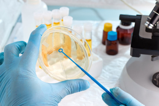
Two new papers appeared recently in the journal Cell, investigating how SARS-CoV-2 infects cells.

So, how exactly does the virus gain entry to cells, and why is it important to know this?

Understanding the target molecules that facilitate viral entry into cells is paramount to identifying how to stop this process from happening.

Both papers report that SARS-CoV-2 makes use of the same mechanism for viral entry that the SARS virus (SARS-CoV) uses.

More importantly, both research teams looked at ways of disrupting this process, using an enzyme inhibitor and antibodies against the SARS virus.

Coronavirus infection route
The new coronavirus, SARS-CoV-2, is a type of virus called an enveloped RNA virus.

This means that its genetic material is encoded in single-stranded RNA molecules surrounded by a cell membrane taken from the cell that it last infected.

When enveloped viruses infect a cell, they do this using a two-stage process.

The first step involves making a connection with a receptor on the surface of the target cell. The second is fusion with a cell membrane, either on the surface of the cell or at an internal location.

In the case of coronaviruses, the first step requires that specific proteins in the viral envelope, called spike (S) proteins, undergo a biochemical modification. This step is called S protein priming.

The enzymes responsible for S protein priming are potential therapeutic targets as inhibiting their mechanism may prevent a virus from being able to enter a cell.

“Unravelling which cellular factors are used by SARS-CoV-2 for entry might provide insights into viral transmission and reveal therapeutic targets,” write the authors one of the new papers in Cell.

The senior study author is Stefan Pöhlmann, a professor for Infection Biology at Georg-August-University and Head of the Infection Biology Unit of the German Primate Center, both in Göttingen in Germany.

Pöhlmann and his colleagues show evidence that the SARS-CoV-2 S protein binds to the same receptor as the SARS virus S protein. The receptor is called angiotensin-converting enzyme 2 or ACE2.

In fact, an earlier paper in the journal Nature had already implicated ACE2 as the receptor that allows SARS-CoV-2 to infect cells.

In addition to providing further evidence of ACE2’s role, Pöhlmann and the team also saw that, like SARS-CoV, the new coronavirus S protein uses an enzyme called TMPRSS2 for S protein priming.

Importantly, they showed that “camostat mesylate, an inhibitor of TMPRSS2, blocks SARS-CoV-2 infection of lung cells.”

Camostat mesylate is a drug approved in Japan for the treatment of pancreatitis. The authors explain in the paper:

Towards a SARS-CoV-2 vaccine
Pöhlmann and his colleagues also studied whether antibodies made by people who had a previous diagnosis of SARS would prevent SARS-CoV-2 virus entry into cells.

They found that antibodies against the SARS-CoV S protein reduced how well a laboratory model virus with the SARS-CoV-2 S protein could infect cells. They also saw similar results with antibodies against S proteins made in rabbits.

“Although confirmation with infectious virus is pending, our results indicate that neutralizing antibody responses raised against SARS-S could offer some protection against SARS-CoV-2 infection, which may have implications for outbreak control,” the team writes in the paper.

Yet, Pöhlmann and his colleagues are not the only ones studying the potential to use antibodies to SARS as a vaccine for SARS-CoV-2.

David Veesler, an assistant professor in Biochemistry at the University of Washington in Seattle, provides more evidence that the virus enters target cells via ACE2 in a paper published in Cell.

Along with his colleagues, he also studied antibodies against SARS S protein fragments to identify potential vaccines.

The team showed that antibody serum from four different mice could reduce infection with a laboratory model virus containing the SARS-CoV-2 S by 90%.

But before a much-needed SARS-CoV-2 vaccine is available, more testing is required.

Clinical trials to show the safety and efficacy will form the basis of developing these vaccine candidates into safe products to use.

In Europe, the European Medicines Agency announced last month that it was taking “concrete actions to accelerate the development and availability of medicinal products for the treatment and prevention of the new coronavirus.”

Meanwhile, in the United States, the Department of Health and Human Services is collaborating with Janssen Research and Development, part of pharmaceutical company Johnson & Johnson, to develop a vaccine against SARS-CoV-2. A clinical trial, sponsored by the National Institute of Allergy and Infectious Diseases using a novel type of RNA-based vaccine, is also underway.

Antibiotics discovered that kill bacteria in a new way

A new group of antibiotics with a unique approach to attacking bacteria has been discovered, making it a promising clinical candidate in the fight against antimicrobial resistance.

The newly-found corbomycin and the lesser-known complestatin have a never-before-seen way to kill bacteria, which is achieved by blocking the function of the bacterial cell wall. The discovery comes from a family of antibiotics called glycopeptides that are produced by soil bacteria.

The researchers also demonstrated in mice that these new antibiotics can block infections caused by the drug resistant Staphylococcus aureus which is a group of bacteria that can cause many serious infections.

The findings were published in Nature today.

“Bacteria have a wall around the outside of their cells that gives them shape and is a source of strength,” said study first author Beth Culp, a PhD candidate in biochemistry and biomedical sciences at McMaster.

“Antibiotics like penicillin kill bacteria by preventing building of the wall, but the antibiotics that we found actually work by doing the opposite — they prevent the wall from being broken down. This is critical for cell to divide.

“In order for a cell to grow, it has to divide and expand. If you completely block the breakdown of the wall, it is like it is trapped in a prison, and can’t expand or grow.”

Looking at the family tree of known members of the glycopeptides, researchers studied the genes of those lacking known resistance mechanisms, with the idea they might be an antibiotic demonstrating a different way to attack bacteria.

“We hypothesized that if the genes that made these antibiotics were different, maybe the way they killed the bacteria was also different,” said Culp.

The group confirmed that the bacterial wall was the site of action of these new antibiotics using cell imaging techniques in collaboration with Yves Brun and his team from the Université de Montréal.

Culp said: “This approach can be applied to other antibiotics and help us discover new ones with different mechanisms of action. We found one completely new antibiotic in this study, but since then, we’ve found a few others in the same family that have this same new mechanism.”

The team is led by professor Gerry Wright of the David Braley Centre for Antibiotic Discovery within the Michael G. DeGroote Institute for Infectious Disease Research at McMaster.

The research was funded by the Canadian Institutes of Health Research and the Ontario Research Fund.

Scientists breach brain barriers to attack tumors

The brain is a sort of fortress, equipped with barriers designed to keep out dangerous pathogens. But protection comes at a cost: These barriers interfere with the immune system when faced with dire threats such glioblastoma, a deadly brain tumor for which there are few effective treatments.

Yale researchers have found a novel way to circumvent the brain’s natural defenses when they’re counterproductive by slipping immune system rescuers through the fortresses’ drainage system, they report Jan. 15 in the journal Nature.

“People had thought there was very little the immune system could do to combat brain tumors,” said senior corresponding author Akiko Iwasaki. “There has been no way for glioblastoma patients to benefit from immunotherapy.”

Iwasaki is the Waldemar Von Zedtwitz Professor of Immunobiology and professor of molecular, cellular, and developmental biology and an investigator for the Howard Hughes Medical Institute.

While the brain itself has no direct way for disposing of cellular waste, tiny vessels lining the interior of the skull collect tissue waste and dispose of it through the body’s lymphatic system, which filters toxins and waste from the body. It is this disposal system that researchers exploited in the new study.

These vessels form shortly after birth, spurred in part by the gene known as vascular endothelial growth factor C, or VEGF-C.

Yale’s Jean-Leon Thomas, associate professor of neurology at Yale and senior co-corresponding author of the paper, wondered whether VEGF-C might increase immune response if lymphatic drainage was increased. And lead author Eric Song, a student working in Iwasaki’s lab, wanted to see if VEGF-C could specifically be used to increase the immune system’s surveillance of glioblastoma tumors. Together, the team investigated whether introducing VEGF-C through this drainage system would specifically target brain tumors.

The team introduced VEGF C into the cerebrospinal fluid of mice with glioblastoma and observed an increased level of T cell response to tumors in the brain. When combined with immune system checkpoint inhibitors commonly used in immunotherapy, the VEGF-C treatment significantly extended survival of the mice. In other words, the introduction of VEGF-C, in conjunction with cancer immunotherapy drugs, was apparently sufficient to target brain tumors.

“These results are remarkable,” Iwasaki said. “We would like to bring this treatment to glioblastoma patients. The prognosis with current therapies of surgery and chemotherapy is still so bleak.”

The study was primarily funded by the Howard Hughes Medical Institute and the National Institutes of Health.

Other Yale authors are Tianyang Mao, Huiping Dong, Ligia Simoes,Braga Boisserand, and Marcus Bosenberg. Salli Antila and Kari Alitalo of the University of Helsinki are also authors.

Revolutionising radiotherapy: making a cornerstone cancer treatment more personal and powerful

“We have a very powerful treatment which contributes to the cure of cancer in around a third of patients treated,” says Professor David Sebag-Montefiore from the University of Leeds of radiotherapy, a cornerstone of cancer treatment in the UK.

But there’s always room for improvement. “Some of the radiotherapy we give today isn’t doing a good enough job.”

We want more people to reap the benefits of this tried and tested treatment, so we’re investing £56 million to launch the Cancer Research UK Radiation Research Network (CRUK RadNet). This will support radiotherapy research in seven specialist institutes across the UK and aims to propel radiotherapy into the future.

It’s money to develop new tech, harness the power of existing ones, apply approaches like artificial intelligence (AI) and to help scientists really understand what’s going on when cancer cells are hit by radiotherapy beams, so we can use drugs to boost their cancer-killing effects.

“Because radiotherapy is an effective treatment across such a broad range of cancers it’s clearly a job that can’t be achieved in one centre alone,” says Sebag-Montefiore. This new initiative hopes to make use of the different expertise from each research station involved, bringing in knowledge from scientists who have never worked in radiotherapy before. “This network will allow us to focus along the breath of cancer research and actually make a big impact.”

Although radiotherapy has become extremely advanced in the last couple of decades and comes in many high-tech forms, there are still plenty of questions around how it’s effects can be maximised to benefit more people and how to reduce side effects of the treatment.

Here are just three of a number of research areas CRUK RadNet hopes to get answers for.

How does radiotherapy affect the tumour microenvironment?

Radiotherapy works by damaging the DNA of cancer cells. “These fatal DNA breaks mean the cancer cells can’t divide,” says Sebag-Montefiore.

But cancer cells aren’t just quietly hanging out by themselves. They’re sitting in a busy environment of blood cells, immune cells and healthy tissues, all of whom are likely to be interacting.

The full extent of the influence these cells have on each other is still unclear, but clues from the clinic suggest that the immune system plays an important role in mopping up cancer cells after radiotherapy, even after they’ve spread to other parts of the body.

“Clinically we are now starting to see situations in people who have incurable cancer that has spread, where irradiating the primary tumour improves their survival,” says Sebag-Montefiore. It’s a positive benefit that scientists are still working to understand.

One theory is that radiation causes cancer cells to break down and release their contents, which creates lots of interesting molecules for the immune system to detect and home in on. The energised immune cells then move around the body looking for cancer cells displaying these same molecules. And when they find them, they kill them, which may explain why tumours that are distant from where the cancer first started shrink.

Figuring out what’s going on in and around a tumour when it’s being irradiated could point to ways to enhance treatment and may even help radiotherapy work for those who currently don’t respond to it. For example, immunotherapies could give immune cells the boost they need to turn against tumours. Or, giving patients drugs that stop cancer cells repairing their DNA alongside radiotherapy might give the cancer an extra blow.

Sebag-Montefiore says there is clearly a substantial piece of work needed to understand the environment that the radiotherapy beams operate in and are also creating. Learning more about this will help us “know how to best to harness the microenvironment and have maximum impact”.

“This is a significant part of the work that will be done in CRUK RadNet.”

How could AI improve radiotherapy?

Every day we hear of new ways that AI is improving everything from how we shop to healthcare. Now Sebag-Montefiore and his team in Leeds, alongside researchers across the CRUK RadNet network, are exploring how it can be used to help cancer treatment.

“In Leeds, we think we can harness AI to actually improve how radiotherapy is given to patients.”

Computer programmes may help them deliver a highly targeted radiotherapy called SABR more accurately, sparing healthy cells. “Both the tumour and the organ we target during treatment move so we apply a safety margin around the tumour to make sure we hit the whole tumour. At the moment this margin is pretty much the same for every patient.”

But the money from CRUK RadNet will allow them to develop algorithms that could work out the exact area the radiotherapy beam will need to cover for each patient. The team hope to use AI to analyse MRI scans from a range of cancers that are in areas of the body that move a lot, like the liver.

“We could analyse those scans with AI to build up a personalised picture of the actual movement,” which means more precise treatment and fewer side effects.

Why do cancer cells become resistant to radiotherapy?

Unfortunately, radiotherapy doesn’t work on everyone.

“We need to understand why radiotherapy isn’t as effective as it could be in some patients,” says Sebag-Montefiore.

“Within all cancers we can identify some groups of patients where radiotherapy resistance is a major barrier to the cure of cancer.”

This may be someone whose cancer initially responds well to treatment but then it stops working. Or, in a few cases, radiotherapy might have no affect at all.

CRUK RadNet members are investigating resistance from a number of angles, looking at ways to overcome the fact that radiotherapy can’t destroy certain cancers. They’re particularly focusing on the cancer types where survival is still depressingly low, like pancreatic cancer and brain tumours.

For instance, a team in Manchester are delving into how radiotherapy changes the biology of cancer cells and how this may contribute to them standing firm against its powerful beams.

A community effort

Sebag-Montefiore is keen to get going. “The last 10 years have seen significant progress in radiotherapy research, but we can do better.”

He says the network now needs to make sure they work with the whole UK radiotherapy community to make sure they do the best science. Because for Sebag-Montefiore, the potential impact of the network is huge.

“CRUK RadNet is a fantastic investment because it gives us a great chance in improving radiotherapy cure rates and reduce side effects further to ensure patients are getting the best possible treatment.”

Good noise, bad noise: White noise improves hearing

Noise is not the same as noise — and even a quiet environment does not have the same effect as white noise. With a background of continuous white noise, hearing pure sounds becomes even more precise, as researchers from the University of Basel have shown in a study in Cell Reports. Their findings could be applied to the further development of cochlear implants.

Despite the importance of hearing in human communication, we still understand very little of how acoustic signals are perceived and how they are processed to allow us to make sense of them. One thing is clear though: the more precisely we can distinguish sound patterns, the better our hearing is. But how does the brain manage to distinguish between relevant and less relevant information — especially in an environment with background noise?

Exploring the “auditory brain”

Researchers led by Prof. Dr. Tania Rinaldi Barkat from the Department of Biomedicine at the University of Basel have investigated the neuronal foundation of sound perception and sound discrimination in a challenging sound environment. The focus was on research into the auditory cortex — the “auditory brain,” that is, the area of the brain that processes acoustic stimuli. The resulting activity patterns stem from measurements in a mouse brain.

As is well known, the distinction between sounds becomes more difficult the closer they are in the frequency spectrum. Initially, the researchers assumed that additional noise could make such a hearing task even more difficult. However, the opposite was observed: The team was able to demonstrate that the brain’s ability to distinguish subtle tone differences improved when white noise was added to the background. Compared to a quiet environment, the noise thus facilitated auditory perception.

Noise reduces neuronal activity

The data of the research group showed that white noise significantly inhibited the activity of the nerve cells in the auditory cortex. Paradoxically, this suppression of the neuronal excitation led to a more precise perception of the pure tones. “We found that less overlap occurred between populations of neurons during two separate tone representations,” explains Professor Tania Barkat. “As a result, the overall reduction in neuronal activity produced a more distinct tone representation.”

To confirm that the auditory cortex and not another area of the brain was responsible for the change in sound perception, the researchers used the light-controlled technique optogenetic. Their findings could possibly be used to improve auditory perception in situations where sounds are difficult to distinguish. According to Barkat, it is conceivable that cochlear implants could be stimulated with an effect similar to white noise in order to improve the frequency resolution and thus the hearing result of their users.

Measuring up: how does the UK compare internationally on cancer survival?

Our ambition is that 3 in 4 people will survive their cancer by 2034. And while cancer outcomes may differ from country to country, the goal of improving cancer survival is one that’s reflected across the world.

A good way for countries to monitor their progress in improving cancer care is by comparing how many people get cancer (incidence), how many survive (survival) and how many die from their cancer (mortality) to see how they measure up. If survival is higher, and incidence and mortality are lower, it’s clear that a country is on the right track.

“No one country manages cancer perfectly,” says John Butler, a consultant specialising in gynaecological cancer surgery. “ But international studies enable countries to learn lessons from one another to with the aim of improving their own cancer policies.”

And in the latest study, published in Lancet Oncology by the International Cancer Benchmarking Partnership, some promising trends have emerged. Survival has improved for the 7 cancer types studied in all countries between 1995 and 2014.

But the figures also underline how much progress still needs to be made in the UK to equal the best outcomes globally. With the exception of ovarian and oesophageal, the UK has the lowest survival figures for the cancers studied.

What do the latest figures show?

Big, international studies like this are a task for the International Cancer Benchmarking Partnership (ICBP). Led by clinicians, researchers and policymakers from around the world, the team compare trends in cancer survival, incidence and mortality rates across 7 countries with similar healthcare systems: UK, Australia, Canada, New Zealand, Denmark, Norway and Ireland. Something that’s never been done before.

Comparisons like this can be tricky – mainly because countries collect and record data in slightly different ways, something the ICBP is looking at in more detail. But despite the challenges, the latest figures from the ICBP are the best available and will only get better as more analysis is done.

The team has been collecting data from 7 cancer types – ovary, lung, colon, rectum, pancreas, oesophagus and stomach – since 1995.

And the latest figures, covering 1995 to 2014, reveal some stark differences in cancer survival between countries. Generally, cancer survival is higher in Australia, Canada and Norway than in Denmark, Ireland, New Zealand and the UK.

Similar trends can be seen for individual cancer types, like lung cancer. From the graphs we can see that Australia has the highest lung cancer survival, and Ireland has made the greatest increase in survival over time. But despite big improvements in lung cancer survival, the UK remains bottom of the list for this cancer type.

Why is the UK lagging behind?

There are many, complex reasons that could explain why we have lower survival compared to other countries.

Butler, the lead clinical advisor for ICBP, says there are some factors that will affect survival in all cancer types. “The UK health system is under great pressure, with increasing demands on cancer diagnostics and more urgent referrals”. And that could affect survival figures. Diagnosing and treating cancer early gives patients the best chance of surviving their cancer, but it relies on having enough NHS staff and funding in place to make this a reality – something the NHS doesn’t currently have.

But there are also more specific reasons that may explain differences between countries for some cancers.
Take ovarian cancer for example. Patients diagnosed in the UK appear to be diagnosed at similar stages to other countries, but survival is lower. This suggests there could be improvements in how these patients are treated.

And as Butler elaborated, this is amplified in older patients.

Older patients are more likely to have other health problems, which often make it more challenging to perform surgery or deliver chemotherapy. More is needed to be done to understand these patients’ complex needs and improve treatments for them, as we’ve blogged about before, as well as to understand why this is an issue particularly for the UK.

But when looking at survival as a whole, it’s useful to consider where we started. In 1995, the UK had some of the lowest survival estimates of the seven countries studied. This means that even though we have made improvements in certain cancers, we’re starting from a lower baseline. Which makes it that much harder for us to catch up with the countries who have higher survival.

And it’s where comparing our progress to other countries can help.

What can we learn from other countries?

Denmark was in a similar place to the UK with their survival in 1995. But as Jesper Fisker, chief executive officer of the Danish Cancer Society, told us “There’s been great progress in Danish cancer survival – which is the result of massive efforts and investments in the cancer field over the past 20 years”.

They’ve also made major strides towards centralising their cancer services, meaning cancer patients are treated in fewer, more specialised centres, with the best clinicians for their cancer type.

And it’s paid off – Denmark has seen real improvements in cancer survival – such as increasing their 1-year survival of lung cancer from 27.5% to 46.2% (from 1995-1999 to 2010-2014). The UK has made similar efforts to improve cancer services, with some success, but much more needs to be done.

And it’s not quite as straightforward as it sounds. While Denmark has made big improvements overall, this has not been universal for every cancer type. The same is true for all the countries studied and it’s something the ICBP is working to understand. They’re looking into variations in access people have to diagnostic tests, scans and treatment, as well as differences in healthcare systems that could help to explain the disparity.

Progress for the UK

On the bright side, the UK has made particularly good progress in increasing cancer survival in rectal, ovarian, and oesophageal cancers.

For example, from 1995 to 1999, 48 in 100 patients were estimated to survive their rectal cancer for at least 5 years. This has now increased to 62 in 100 patients for 2010-2014, only 8.7% behind Australia, who had the highest rectal cancer survival of the countries studied.

Butler called the progress “encouraging” and said there were lots of factors that could be behind the improvements. England produced its first national cancer plan in 2000 and appointed a national cancer director, who helps provide advice and leadership for our cancer services. Since then there’s been more guidance and greater scrutiny of how cancer services are performing, as well as more funding.

There’s also been a move towards cancers being treated in specialised centres, where there will be more relevant cancer expertise.

How can the UK catch up?

But despite the improvements, there’s clearly more work to be done in the UK.

For Butler, investigations into how well cancer services are performing could be a good way to start. For example, national audits in the UK for lung cancer have increased the number of people having surgery, as well as the number of specialised lung cancer surgeons. Replicating this approach could help the NHS direct its efforts to improve outcomes for other types of cancer.

And while the UK government have introduced a range of policies between 1995 and 2014 to improve cancer services and speed up diagnosis and treatment, these have added to the strain on NHS services.

It’s crucial that investment into cancer services is increased to match the ever-growing demand. As Butler told us, “one of the biggest challenges the UK faces is capacity of diagnostic services.”

Trump signs order aimed at development of better flu vaccines

Reporting By Deena Beasley; Editing by Sonya Hepinstall

(Reuters) – U.S. President Donald Trump on Thursday signed an executive order aimed at spurring the development of better vaccines to protect against seasonal influenza as well as a potential pandemic flu outbreak.

The order does not allocate additional funding for now, but calls for an evaluation of current flu vaccine manufacturing abilities and a task force report including cost estimates, administration officials said on a call with reporters.
Each year the seasonal flu, which can kill tens of thousands of Americans, costs the United States about $50 billion, including lost productivity, one administration official said. A serious flu pandemic would push those costs to between $1.8 billion and $3.8 billion, he said.

Manufacturers such as GlaxoSmithKline Plc and Sanofi SA make millions of doses of flu vaccine for the U.S. market alone, growing the virus in chicken eggs. Usually the doses, which protect against strains that experts predicted the previous February, are ready in time and in sufficient quantity for the winter flu season.
But if the strain that appears during flu season was not the one experts forecast, the vaccines might not work. The appearance of H1N1 swine flu in 2009-2010 took experts by surprise, and the flu was already on its second wave before a vaccine was ready; an estimated 61 million people in the U.S. got swine flu and thousands died.

The Department of Health and Human Services will coordinate government efforts to modernize influenza vaccine production. The focus will be on recombinant technologies to quickly produce reliable vaccines as well as “universal” vaccines that would elicit immunity against parts of the virus that do not change from year to year.

NHS to fund ‘life changing’ haemophilia drug

Mark Gould

Thursday, 22 August 2019

NHS England is to fund what it says will be “life changing treatment” for around 2,000 people with severe Haemophilia A, which will dramatically cut their risk of life-threatening bleeds and reduce treatment time.

The new treatment is part of a package of measures set out in the NHS Long Term Plan which will save lives through access to the most advanced medical interventions.

A new drug – emicizumab (also referred to as Hemlibra®) – mimics the action of the blood protein factor VIII to avoid uncontrolled bleeding, while cutting treatment times from multiple time-consuming infusions every week to a single injection given once-a-week or fortnight.

Many young children are affected by the condition and NHS England says parents sometimes struggle to administer the current infusion several times a month.

Simon Stevens, NHS chief executive, said: “Giving patients access to world class, trailblazing drugs and therapies is a key part of the NHS Long Term Plan which aims to save thousands more lives.

“As a parent I know that cuts and scrapes happen to kids all the time, but for many families these routine accidents can be distressing and life-threatening, so this new treatment will change lives and lift a weight from thousands of parents.

“This treatment has the potential to significantly improve the lives of people with haemophilia, especially children – reducing treatment time and even ending the dangerous bleeds which can lead to life-threatening cuts and life-changing damage.”

Liz Carroll, chief executive of The Haemophilia Society, said: “This decision is fantastic news for our community. Current treatments can require intravenous infusions multiple times a week which can place a significant burden on people with haemophilia and their carers. This decision will mean that people will have the opportunity to have treatment less frequently without intravenous access which will enable many to live their lives more freely.’’

Gene mutation evolved to cope with modern high-sugar diets

The gene variant became more common in humans after cooking and farming became widespread, and might now help people avoid diabetes, according to the findings published in eLife.

“We found that people differ in how efficiently their bodies can manage blood sugar levels, resulting from an evolutionary process that seems to have been brought about by changing diets,” said the study’s lead author, Professor Frances Brodsky, Director of UCL Biosciences.

The researchers were investigating the CLTCL1 gene, which directs production of the CHC22 protein that plays a key role in regulating a glucose transporter in our fat and muscle cells.

After people eat, the hormone insulin reacts to higher levels of blood glucose by releasing the transporter to remove glucose from the blood, taking it into muscle and fat tissue. Between meals, with the help of the CHC22 protein, the glucose transporter remains inside muscle and fat so that some blood sugar will continue to circulate.

The research team, consisting of specialists in population genetics, evolutionary biology, ancient DNA and cell biology, analysed human genomes as well as those of 61 other species, to understand how the gene producing CHC22 has varied throughout evolutionary history.

In humans, by looking at the genomes of 2,504 people from the global 1000 Genomes Project, they found that almost half of the people in many ethnic groups have a variant of CHC22 that is produced by a mutated gene, which became more common as people developed cooking and farming.

The researchers also looked at genomes of ancient humans, and found that the newer variant is more common in ancient and modern farming populations than in hunter-gatherers, suggesting that increased consumption of carbohydrates could have been the selective force driving the genetic adaptation.

By studying cells, the researchers found that the newer CHC22 variant is less effective at keeping the glucose transporter inside muscle and fat between meals, meaning the transporter can more readily clear glucose out of the blood. People with the newer variant will therefore have lower blood sugar.

“The older version of this genetic variant likely would have been helpful to our ancestors as it would have helped maintain higher levels of blood sugar during periods of fasting, in times when we didn’t have such easy access to carbohydrates, and this would have helped us evolve our large brains,” said first author Dr Matteo Fumagalli, who began the study at UCL before moving to Imperial College London.

“In more recent years, with our high-carb diets that often provide us too much sugar, the newer variant may be advantageous,” Dr Fumagalli added.

The researchers say that while this genetic variant does not play a direct role in the development of diabetes, having the older variant may make people more likely to develop diabetes, and it may also exacerbate insulin resistance involved in diabetes.

“People with the older variant may need to be more careful of their carb intake, but more research is needed to understand how the genetic variant we found can impact our physiology,” added Professor Brodsky.

Co-author Professor Mark Thomas (UCL Genetics, Evolution & Environment) added: “Our analyses strongly suggest that we have found yet another example of how prehistoric changes in dietary habits have shaped human evolution. Understanding how we have adapted to these changes doesn’t only inform us about why people lived or died in the past, but also helps us to better understand the relationship between diet, health and disease today.”

The study was funded by the National Institutes of Health (USA), Wellcome and the Medical Research Council (UK).

Genetic therapy heals damage caused by heart attack

Researchers from King’s College London have found that therapy that can induce heart cells to regenerate after a heart attack.

Myocardial infarction, more commonly known as a heart attack, caused by the sudden blocking of one of the cardiac coronary arteries, is the main cause of heart failure, a condition that now affects over 23 million population in the world, according to the World Health Organisation.

At present, when a patient survives a heart attack, they are left with permanent structural damage to their heart through the formation of a scar, which can lead to heart failure in the future. In contrast to fish and salamander, which can regenerate the heart throughout life.

In this study, published today in Nature, the team of investigators delivered a small piece of genetic material, called microRNA-199, to the heart of pigs, after a myocardial infarction which resulted in the almost complete recovery of cardiac function at one month later.

Lead author Professor Mauro Giacca, from King’s College London said: “It is a very exciting moment for the field. After so many unsuccessful attempts at regenerating the heart using stem cells, which all have failed so far, for the first time we see real cardiac repair in a large animal.”

This is the first demonstration that cardiac regeneration can be achieved by administering an effective genetic drug that stimulates cardiac regeneration in a large animal, with heart anatomy and physiology like that of humans.

“It will take some time before we can proceed to clinical trials” explained Professor Giacca.

“We still need to learn how to administer the RNA as a synthetic molecule in large animals and then in patients, but we already know this works well in mice.”