Can a dangerous microbe offer a new way to silence pain?

Can a Dangerous Microbe Offer a New Way to Silence Pain? - Neuroscience News

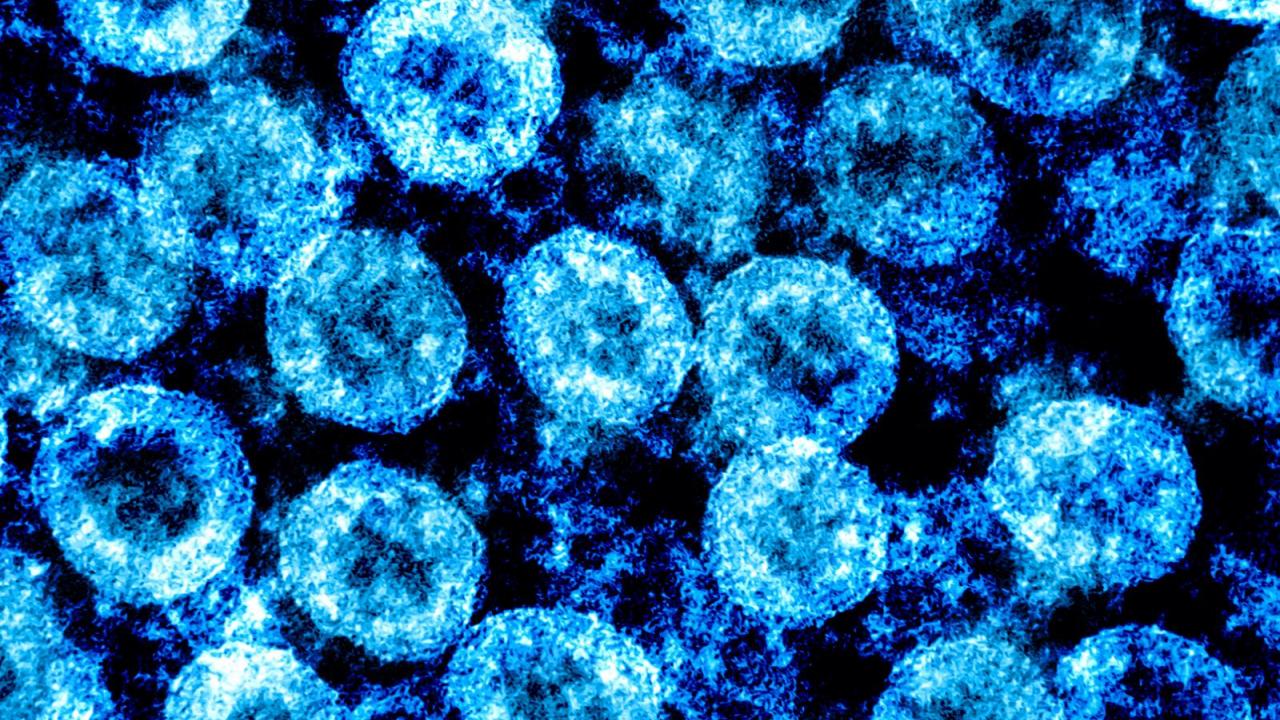
Anthrax has a scary reputation. Widely known to cause serious lung infections in humans and unsightly, albeit painless, skin lesions in livestock and people, the anthrax bacterium has even been used as a weapon of terror.

Now the findings of a new study suggest the dreaded microbe also has unexpected beneficial potential — one of its toxins can silence multiple types of pain in animals.

The research reveals that this specific anthrax toxin works to alter signaling in pain-sensing neurons and, when delivered in a targeted manner into neurons of the central and peripheral nervous system, can offer relief to animals in distress.

The work, led by investigators at Harvard Medical School in collaboration with industry scientists and researchers from other institutions, is published Dec. 20 in Nature Neuroscience.

Furthermore, the team combined parts of the anthrax toxin with different types of molecular cargo and delivered it into pain-sensing neurons. The technique can be used to design novel precision-targeted pain treatments that act on pain receptors but without the widespread systemic effects of current pain-relief drugs, such as opioids.

“This molecular platform of using a bacterial toxin to deliver substances into neurons and modulate their function represents a new way to target pain-mediating neurons,” said study senior investigator Isaac Chiu, associate professor of immunology in the Blavatnik Institute at Harvard Medical School.

The need to expand the current therapeutic arsenal for pain management remains acute, the researchers said. Opioids remain the most effective pain medication, but they have dangerous side effects — most notably their ability to rewire the brain’s reward system, which makes them highly addictive, and their propensity to suppress breathing, which can be fatal.

“There’s still a great clinical need for developing non-opioid pain therapies that are not addictive but that are effective in silencing pain,” said study first author Nicole Yang, HMS research fellow in immunology in the Chiu Lab. “Our experiments show that one strategy, at least experimentally, could be to specifically target pain neurons using this bacterial toxin.”

The researchers caution, however, that for now, this approach remains purely experimental and still needs to be tested and further fine-tuned in more animal studies and, eventually, in humans.

Primed to connect

Researchers in the Chiu lab have long been interested in the interplay between microbes and the nervous and immune systems. Past work led by Chiu has demonstrated that other disease-causing bacteria can also interact with neurons and alter their signaling to amplify pain. Yet only a handful of studies so far have looked at whether certain microbes could minimize or block pain. This is what Chiu and Yang set out to do.

For the current study, they started out by trying to determine how pain-sensing neurons may be different from other neurons in the human body. To do so, they first turned to gene-expression data. One of the things that caught their attention: Pain fibers had receptors for anthrax toxins, whereas other types of neurons did not. In other words, the pain fibers were structurally primed to interact with the anthrax bacterium. They wondered why.

The newly published research sheds light on that very question.

The findings demonstrate that pain silencing occurs when sensory neurons of dorsal root ganglia, nerves that relay pain signals to the spinal cord, connect with two specific proteins made by the anthrax bacterium itself. Experiments revealed that this occurs when one of the bacterial proteins, protective antigen (PA), binds to the nerve cell receptors it forms a pore that serves as a gateway for two others bacterial proteins, edema factor (EF) and lethal factor (LF), to be ferried into the nerve cell. The research further demonstrated PA and EF together, collectively known as edema toxin, alter the signaling inside nerve cells — in effect silencing pain.

Using the quirks of microbial evolution for new therapies

In a series of experiments, the researchers found that the anthrax toxin altered signaling in human nerve cells in dishes, and it also did so in living animals.

Injecting the toxin into the lower spines of mice produced potent pain-blocking effects, preventing the animals from sensing high-temperature and mechanical stimulations. Importantly, the animals’ other vital signs such as heart rate, body temperature, and motor coordination were not affected — an observation that underscored that this technique was highly selective and precise in targeting pain fibers and blocking pain without widespread systemic effects.

Furthermore, injecting mice with the anthrax toxin alleviated symptoms of two other types of pain: pain caused by inflammation and pain caused by nerve cell damage, often seen in the aftermath of traumatic injury and certain viral infections such as herpes zoster, or shingles, or as a complication of diabetes and cancer treatment.

Additionally, the researchers observed that as the pain diminished, the treated nerve cells remained physiologically intact — a finding that indicates the pain-blocking effects were not due to injury of the nerve cells but rather stemmed from the altered signaling inside them.

In a final step, the team designed a carrier vehicle from anthrax proteins and used it to deliver other pain-blocking substances into nerve cells. One of these substances was botulinum toxin, yet another potentially lethal bacterium known for its ability to alter nerve signaling. That approach, too, blocked pain in mice. The experiments demonstrate this could be a novel delivery system for targeting pain.

“We took parts of the anthrax toxin and fused them to the protein cargo that we wanted it to deliver,” Yang said. “In the future, one could think of different kinds of proteins to deliver targeted treatments.”

The scientists caution that as the work progresses, the safety of the toxin treatment must be monitored carefully, especially given that the anthrax protein has been implicated in disrupting the integrity of the blood-brain barrier during infection.

The new findings raise another interesting question: Evolutionarily speaking, why would a microbe silence pain?

Chiu thinks that one explanation — a highly speculative one, he added — may be that microbes have developed ways to interact with their host in order to facilitate their own spread and survival. In the case of anthrax, that adaptive mechanism may be through altered signaling that blocks the host’s ability to sense pain and therefore the microbe’s presence. This hypothesis could help explain why the black skin lesions that the anthrax bacterium sometimes forms are notably painless, Chiu added.

The new findings also point to novel avenues for drug development beyond the traditional small-molecule therapies that are currently being designed across labs.

“Bringing a bacterial therapeutic to treat pain raises the question ‘Can we mine the natural world and the microbial world for analgesics?'” Chiu said. “Doing so can increase the range and diversity of the types of substances we look to in search for solutions.”

Coinvestigators included Jörg Isensee, Dylan Neel, Andreza Quadros, Han-Xiong Bear Zhang, Justas Lauzadis, Sai Man Liu, Stephanie Shiers, Andreea Belu, Shilpa Palan, Sandra Marlin, Jacquie Maignel, Angela Kennedy- Curran, Victoria Tong, Mahtab Moayeri, Pascal Röderer, Anja Nitzsche, Mike Lu, Bradley Pentelute, Oliver Brüstle, Vineeta Tripathi, Keith Foster, Theodore Price, John Collier, Stephen Leppla, Michelino Puopolo, Bruce Bean, Thiago Cunha, and Tim Hucho.

This study was funded by the Burroughs Wellcome Fund; Chan-Zuckerberg Initiative; Ipsen Pharmaceuticals; National Institutes of Health (DP2AT009499, R01AI130019, R01NS036855, NIA 5T32AG000222 fellowship, NIH NIGMS T32GM007753 fellowship), and NIH NINDS (NS111929); National Institute of Allergy and Infectious Diseases Intramural Program; European Regional Development Fund (NeuRoWeg, EFRE?0800407 and EFRE?0800408); Innovative Medicines Initiative 2 Joint Undertaking (116072-NGN-PET); and São Paulo Research Foundation (2013/08216-2 Center for Research in Inflammatory Diseases); Deutsche Forschungsgemeinschaft (271522021 and 413120531), EFRE-0800384, and LeitmarktAgentur.NRW (LS-1-1-020d).

Potential role of ‘junk DNA’ sequence in aging, cancer

The human body is essentially made up of trillions of living cells. It ages as its cells age, which happens when those cells eventually stop replicating and dividing. Scientists have long known that genes influence how cells age and how long humans live, but how that works exactly remains unclear. Findings from a new study led by researchers at Washington State University have solved a small piece of that puzzle, bringing scientists one step closer to solving the mystery of aging.

A research team headed by Jiyue Zhu, a professor in the College of Pharmacy and Pharmaceutical Sciences, recently identified a DNA region known as VNTR2-1 that appears to drive the activity of the telomerase gene, which has been shown to prevent aging in certain types of cells. The study was published in the journal Proceedings of the National Academy of Sciences (PNAS).

The telomerase gene controls the activity of the telomerase enzyme, which helps produce telomeres, the caps at the end of each strand of DNA that protect the chromosomes within our cells. In normal cells, the length of telomeres gets a little bit shorter every time cells duplicate their DNA before they divide. When telomeres get too short, cells can no longer reproduce, causing them to age and die. However, in certain cell types — including reproductive cells and cancer cells — the activity of the telomerase gene ensures that telomeres are reset to the same length when DNA is copied. This is essentially what restarts the aging clock in new offspring but is also the reason why cancer cells can continue to multiply and form tumors.

Knowing how the telomerase gene is regulated and activated and why it is only active in certain types of cells could someday be the key to understanding how humans age, as well as how to stop the spread of cancer. That is why Zhu has focused the past 20 years of his career as a scientist solely on the study of this gene.

Zhu said that his team’s latest finding that VNTR2-1 helps to drive the activity of the telomerase gene is especially notable because of the type of DNA sequence it represents.

“Almost 50% of our genome consists of repetitive DNA that does not code for protein,” Zhu said. “These DNA sequences tend to be considered as ‘junk DNA’ or dark matters in our genome, and they are difficult to study. Our study describes that one of those units actually has a function in that it enhances the activity of the telomerase gene.”

Their finding is based on a series of experiments that found that deleting the DNA sequence from cancer cells — both in a human cell line and in mice — caused telomeres to shorten, cells to age, and tumors to stop growing. Subsequently, they conducted a study that looked at the length of the sequence in DNA samples taken from Caucasian and African American centenarians and control participants in the Georgia Centenarian Study, a study that followed a group of people aged 100 or above between 1988 and 2008. The researchers found that the length of the sequence ranged from as short as 53 repeats — or copies — of the DNA to as long as 160 repeats.

“It varies a lot, and our study actually shows that the telomerase gene is more active in people with a longer sequence,” Zhu said.

Since very short sequences were found only in African American participants, they looked more closely at that group and found that there were relatively few centenarians with a short VNTR2-1 sequence as compared to control participants. However, Zhu said it was worth noting that having a shorter sequence does not necessarily mean your lifespan will be shorter, because it means the telomerase gene is less active and your telomere length may be shorter, which could make you less likely to develop cancer.

“Our findings are telling us that this VNTR2-1 sequence contributes to the genetic diversity of how we age and how we get cancer,” Zhu said. “We know that oncogenes — or cancer genes — and tumor suppressor genes don’t account for all the reasons why we get cancer. Our research shows that the picture is a lot more complicated than a mutation of an oncogene and makes a strong case for expanding our research to look more closely at this so-called junk DNA.”

Zhu noted that since African Americans have been in the United States for generations, many of them have Caucasian ancestors from whom they may have inherited some of this sequence. So as a next step, he and his team hope to be able to study the sequence in an African population.

In addition to Zhu, authors on the paper include co-first authors Tao Xu and De Cheng and others at Washington State University, as well as their collaborators at Northeast Forestry University in China; Pennsylvania State University; and North Carolina State University.

Funding for this study came from the National Institutes of Health’s National Institute of General Medical Sciences, the Melanoma Research Alliance, and the Health Sciences and Services Authority of Spokane County.

Novavax COVID-19 vaccine more than 90% effective in U.S. trial

Novavax Inc (NVAX.O) on Monday said its COVID-19 vaccine was more than 90% effective, including against a variety of concerning variants of the coronavirus in a large, late-stage U.S.-based clinical trial.

The study of nearly 30,000 volunteers in the United States and Mexico puts Novavax on track to file for emergency authorization in the United States and elsewhere in the third quarter of 2021, the company said.

The protein-based vaccine was more than 93% effective against the more easily transmissible predominant coronavirus variants that have caused concern among scientists and public health officials, Novavax said.

Protein-based vaccines are a conventional approach that use purified pieces of the virus to spur an immune response, such as those used against whooping cough and shingles.

While the trial was being conducted, the virus variant first discovered in the United Kingdom and now known as the Alpha variant was the most common circulating in the United States, the company said.

The concerning variants first identified in Brazil, South Africa and India were also detected among the trial’s participants, Novavax’s head of research and development, Dr. Gregory Glenn, told Reuters.

The vaccine was 91% effective among volunteers at high risk of severe infection and 100% effective in preventing moderate and severe cases of COVID-19. It was roughly 70% effective against virus variants that Novavax was unable to identify, Glenn said.

“Practically speaking, it’s very important that the vaccine can protect against a virus that is wildly swinging around” in terms of new variants, Glenn said.

Vials labelled “COVID-19 Coronavirus Vaccine” and syringe are seen in front of displayed Novavax logo in this illustration taken, February 9, 2021. REUTERS/Dado Ruvic
Novavax said the vaccine was generally well tolerated. Side effects included headache, fatigue and muscle pain and were generally mild. A small number of participants experienced side effects described as severe.

Novavax remains on track to produce 100 million doses per month by the end of the third quarter of 2021 and 150 million doses per month in the fourth quarter of 2021, the company said.

Chief Executive Stanley Erck told CNBC on Monday it is possible the United States could donate the 110 million shots Novavax has agreed to supply to the U.S. government to the COVAX program that provides COVID-19 vaccines to poorer countries.

The Maryland-based company has repeatedly pushed back production forecasts and has struggled to access raw materials and equipment needed to manufacture its vaccine.

However, in a May investor call, Erck said major manufacturing hurdles have been cleared and that all of its facilities can now produce COVID-19 vaccine at commercial scale.

Erck said Novavax has begun its regulatory filing in India in partnership with the Serum Institute of India (SII), which is contracted to make Novavax shots.

Erck said his understanding is that SII is no longer constrained by raw materials shortages.

SII had said in March that U.S. restrictions on exports of supplies used for vaccines were limiting its ability to scale up production.

Novavax shares were off 3% in midday trading.

Insulin rises before cells develop resistance, new diabetes research implies

Researchers at the University of Gothenburg, Sweden, have now presented results that may change our basic view of how type 2 diabetes occurs. Their study indicates that free fatty acids (FFAs) in the blood trigger insulin release even at a normal blood-sugar level, without an overt uncompensated insulin resistance in fat cells. What is more, the researchers demonstrate the connection with obesity: the amount of FFAs largely depends on how many extra kilos of adipose tissue a person carries, but also on how the body adapt to the increased adiposity.

Worldwide, extensive research is underway to clarify exactly what happens in the body as type 2 diabetes progresses, and why obesity is such a huge risk factor for the disease. For almost 50 years, diabetes researchers have been discussing their version of the chicken-or-egg question: Which comes first — insulin resistance or elevated insulin levels? The dominant hypothesis has long been that the pancreas steps up its insulin production because the cells have already become insulin-resistant, and blood sugar then rises. However, the results now published in the journal EBioMedicine support the opposing idea: that it is the insulin that increases first.

Detailed investigations

The study indicates that high FFA levels in the blood after the overnight fast raise insulin production in the morning. FFAs have long been part of the main research equation for type 2 diabetes, but it is now proposed that they also have another role: in progression of the disease.

For the study, researchers compared metabolism in adipose (fat-storing) tissue among 27 carefully selected research subjects (nine of normal weight, nine with obesity and normal blood sugar, and nine with both obesity and progressed type 2 diabetes). For several days, they underwent extensive examinations in which they had samples taken under varying conditions. The researchers analyzed metabolism and gene expression in the participants’ subcutaneous fat, and the levels of blood sugar, insulin, and FFAs in their blood.

FFAs seem to trigger insulin production

The people with obesity but not diabetes proved to have the same, normal blood-sugar levels as the healthy individuals of normal weight.

“Interestingly, the nondiabetics with obesity had elevated levels of both free fatty acids and insulin in their blood, and those levels were similar to or higher than the levels we were able to measure in blood from the participants with both obesity and type 2 diabetes,” says Emanuel Fryk, resident doctor specializing in general medicine and doctoral student at Sahlgrenska Academy, University of Gothenburg, who is one of the study’s first authors.

In collaboration with researchers at Uppsala University, he observed the same pattern in a population study based on blood samples taken from 500 people after an overnight fast.

“The fact that we saw a link between free fatty acids and insulin there too suggests that the fatty acids are connected with the insulin release, and contribute to increased insulin production on an empty stomach, when blood sugar hasn’t risen,” says Fryk, who nevertheless points out that the finding needs to be confirmed with more research.

Ongoing research

Free fatty acids are found naturally in the bloodstream and, like glycerol, are a product of the body’s fat metabolism. In the subjects, the amount of glycerol released proved to be broadly the same per kilo of body fat, regardless of whether they were of normal weight, had obesity alone, or also had type 2 diabetes.

“Our hypothesis is that the free fatty acids increase in the blood because the adipose tissue can’t store the excess energy anymore. We believe, in that case, it could be an early sign of incipient type 2 diabetes. If our findings are confirmed when other research methods are used, there may be a chance that some specific fatty acids could be developed into biomarkers. But that’s a long way off,” Fryk says.

Lifestyle crucial

Diabetes is one of the most common diseases, with an estimated 500,000 people affected in Sweden. There are also a large number of undetected cases, since many with type 2 diabetes are not yet aware they are ill. Diabetics are at an increased risk for a number of serious conditions, such as cardiovascular disease (which may result in heart attacks and strokes).

“There are many factors that contribute to the progression of type 2 diabetes, but it’s our lifestyle that has, in absolute terms, the largest impact for most people. Our study provides another argument that the most important thing you can do to slow diabetes progression is to change your life style early in the progression of the disease, before blood glucose is elevated, Fryk says.

Study of coronavirus variants predicts virus evolving to escape current vaccines

A new study of the U.K. and South Africa variants of SARS-CoV-2 predicts that current vaccines and certain monoclonal antibodies may be less effective at neutralizing these variants and that the new variants raise the specter that reinfections could be more likely.

The study was published in Nature on March 8, 2021. A preprint of the study was first posted to BioRxiv on January 26, 2021.

The study’s predictions are now being borne out with the first reported results of the Novavax vaccine, says the study’s lead author David Ho, MD. The company reported on Jan. 28 that the vaccine was nearly 90% effective in the company’s U.K. trial, but only 49.4% effective in its South Africa trial, where most cases of COVID-19 are caused by the B.1.351 variant.

“Our study and the new clinical trial data show that the virus is traveling in a direction that is causing it to escape from our current vaccines and therapies that are directed against the viral spike,” says Ho, the director of the Aaron Diamond AIDS Research Center and the Clyde’56 and Helen Wu Professor of Medicine at Columbia University Vagelos College of Physicians and Surgeons.

“If the rampant spread of the virus continues and more critical mutations accumulate, then we may be condemned to chasing after the evolving SARS-CoV-2 continually, as we have long done for influenza virus,” Ho says. “Such considerations require that we stop virus transmission as quickly as is feasible, by redoubling our mitigation measures and by expediting vaccine rollout.”

After vaccination, the immune system responds and makes antibodies that can neutralize the virus.

Ho and his team found that antibodies in blood samples taken from people inoculated with the Moderna or Pfizer vaccine were less effective at neutralizing the two variants, B.1.1.7, which emerged last September in England, and B.1.351, which emerged from South Africa in late 2020. Against the U.K. variant, neutralization dropped by roughly 2-fold, but against the South Africa variant, neutralization dropped by 6.5- to 8.5-fold.

“The approximately 2-fold loss of neutralizing activity against the U.K. variant is unlikely to have an adverse impact due to the large ‘cushion’ of residual neutralizing antibody activity,” Ho says, “and we see that reflected in the Novavax results where the vaccine was 85.6% effective against the U.K. variant.”

Data from Ho’s study about the loss in neutralizing activity against the South Africa variant are more worrisome.

“The drop in neutralizing activity against the South Africa variant is appreciable, and we’re now seeing, based on the Novavax results, that this is causing a reduction in protective efficacy,” Ho says.

The new study did not examine the more recent variant found in Brazil (B.1.1.28) but given the similar spike mutations between the Brazil and South Africa variants, Ho says the Brazil variant should behave similarly to the South Africa variant.

“We have to stop the virus from replicating and that means rolling out vaccine faster and sticking to our mitigation measures like masking and physical distancing. Stopping the spread of the virus will stop the development of further mutations,” Ho says.

The study also found that certain monoclonal antibodies used now to treat COVID patients may not work against the South Africa variant. And based on results with plasma from COVID patients who were infected earlier in the pandemic, the B.1.351 variant from South Africa has the potential to cause reinfection.

New study contains comprehensive analysis of variants

The new study conducted an extensive analysis of mutations in the two SARS-CoV-2 variants compared to other recent studies, which have reported similar findings.

The new study examined all mutations in the spike protein of the two variants. (Vaccines and monoclonal antibody treatments work by recognizing the SARS-CoV-2 spike protein.)

The researchers created SARS-CoV-2 pseudoviruses (viruses that produce the coronavirus spike protein but cannot cause infection) with the eight mutations found in the U.K. variant and the nine mutations found in the South African variant.

They then measured the sensitivity of these pseudoviruses to monoclonal antibodies developed to treat COVID patients, convalescent serum from patients who were infected earlier in the pandemic, and serum from patients who have been vaccinated with the Moderna or Pfizer vaccine.

Implications for monoclonal antibody treatments

The study measured the neutralizing activity of 18 different monoclonal antibodies — including the antibodies in two products authorized for use in the United States.

Against the U.K. variant, most antibodies were still potent, although the neutralizing activity of two antibodies in development was modestly impaired.

Against the South Africa variant, however, the neutralizing activity of four antibodies was completely or markedly abolished. Those antibodies include bamlanivimab (LY-CoV555, approved for use in the United States) that was completely inactive against the South Africa variant, and casirivimab, one of the two antibodies in an approved antibody cocktail (REGN-COV) that was 58-fold less effective at neutralizing the South Africa variant compared to the original virus. The second antibody in the cocktail, imdevimab, retained its neutralizing ability, as did the complete cocktail.

“Decisions of the use of these treatments will depend heavily on the local prevalence of the South Africa and Brazil variants,” Ho says, “highlighting the importance of viral genomic surveillance and proactive development of next-generation antibody therapeutics.”

Reinfection implications

Serum from most patients who had recovered from COVID earlier in the pandemic had 11-fold less neutralizing activity against the South Africa variant and 4-fold less neutralizing activity against the U.K. variant.

“The concern here is that reinfection might be more likely if one is confronted with these variants, particularly the South Africa one,” Ho says.

How do COVID-19 vaccines compare with other existing vaccines?

The novelty of the COVID-19 vaccines may seem daunting for some, and it is natural for questions to arise on their effectiveness. In this feature, we examine the difference between effectiveness and efficacy, compare the COVID-19 frontrunner vaccines to other vaccines, such as the flu shot, and compare their safety considerations.
As Pfizer/BioNTech roll out their COVID-19 vaccine throughout the United Kingdom and the United States, the world wonders how effective it will be.

Looking at the three leading vaccines that we have previously reported on, Pfizer/BioNTech boasts 95% efficacy, the Oxford/AstraZeneca vaccine candidate has an average of 70% efficacy, while the Moderna vaccine candidate reportedly has 94.1% efficacy.

But what does this say about their effectiveness? And how does it compare with vaccines against the flu, polio, and measles?

Effectiveness vs. efficacy — what is the difference?
Firstly, it is worth noting that “effectiveness” and “efficacy” are not the same. Despite news outlets frequently using them interchangeably, efficacy refers to how a vaccine performs under ideal lab conditions, such as those in a clinical trial. In contrast, effectiveness refers to how it performs in the real world.

In other words, in a clinical trial, a 90% efficacy means that there are 90% fewer cases of disease in the group receiving the vaccine compared with the placebo group.

However, the participants chosen for a clinical trial tend to be healthier and younger than those in the general population, and they generally have no underlying conditions. Furthermore, researchers do not normally include certain groups in these studies, such as children or pregnant people.

So, while a vaccine can prevent disease in a trial, we might see this effectiveness drop when administered to the wider population.

However, that is not in itself a bad thing.

Flu shot effectiveness
Vaccines do not need to have high effectiveness to save thousands of lives and prevent millions of disease cases.

The popular flu shot, for example, has an effectiveness of 40­–60%, according to the Centers for Disease Control and Prevention (CDC).

However, during 2018­–2019, it prevented around “4.4 million influenza illnesses, 2.3 million influenza-associated medical visits, 58,000 influenza-associated hospitalizations, and 3,500 influenza-associated deaths.”

It is also worth noting that the flu vaccine’s effectiveness varies from season to season, due to the nature of the flu viruses circulating that year. Determining the precise rate of effectiveness can be challenging.

Finally, it bears mentioning that the number of doses can also improve effectiveness for some vaccines. For the flu shot, two doses of the vaccine instead of one can offer a protection boost, but this benefit is limited to only a few specific groups, such as children or organ transplant recipients.

The booster dose does not seem to benefit people over the age of 65 or those with a compromised immune system.

By contrast, as we will see below, for vaccines, such as the ones against polio and measles, a higher number of doses is required to achieve peak effectiveness.

China coronavirus vaccine may be ready for public in November: official

Coronavirus vaccines being developed in China may be ready for use by the general public as early as November, an official with the Chinese Center for Disease Control and Prevention (CDC) said.

China has four COVID-19 vaccines in the final stage of clinical trials. At least three of those have already been offered to essential workers under an emergency use programme launched in July.

Phase 3 clinical trials were proceeding smoothly and the vaccines could be ready for the general public in November or December, CDC chief biosafety expert Guizhen Wu said in an interview with state TV late on Monday.

Wu, who said she has experienced no abnormal symptoms in recent months after taking an experimental vaccine herself in April, did not specify which vaccines she was referring to.

A unit of state pharmaceutical giant China National Pharmaceutical Group (Sinopharm) and U.S.-listed Sinovac Biotech SVA.O are developing the three vaccines under the state’s emergency use programme. A fourth COVID-19 vaccine being developed by CanSino Biologics 6185.HK was approved for use by the Chinese military in June.

Sinopharm said in July that its vaccine could be ready for public use by the end of this year after the conclusion of Phase 3 trials.

Global vaccine makers are racing to develop an effective vaccine against the virus which has killed more than 925,000 people. Leading Western vaccine makers pledged earlier this month to uphold scientific study standards and reject any political pressure to rush the process.

Cancer Drug Might Help Curb Severe COVID-19

Could a cancer drug spare hospital patients from the ravages of severe COVID-19?

Yale doctors think it can after giving the medication, known as tocilizumab, to severely ill patients back in March.

How does tocilizumab work? It has a long history of dampening the life-threatening immune system reactions cancer patients often experience while undergoing treatment. Since the same kind of dangerous response develops in many COVID-19 cases, the researchers thought the drug might make a difference for the sickest patients.

The result — while preliminary — appears to be a dramatically lower death rate among patients placed on mechanical ventilators.

How much lower? Among the first 239 COVID-19 patients treated at Yale New Haven Hospital, in Connecticut, during the early weeks of the pandemic, 153 were treated with tocilizumab, including all 48 patients who had been placed on ventilators. “Instead of survival rates of 10% to 50% reported elsewhere, it was 75% in [ventilated] patients treated with tocilizumab,” said study author Dr. Christina Price, Yale’s chief of clinical allergy and clinical immunology.

In addition, among those seriously ill patients who ultimately survived COVID-19, tocilizumab appears to have significantly shortened overall ventilation time. While hospitals around the country were having to keep patients hooked up for between 12 to 14 days, ventilations at Yale typically lasted only about five days.

How tocilizumab works against COVID-19

What accounts for its apparent success against COVID-19?

It all originates in the threat posed by a deadly immune system phenomenon known as “cytokine release syndrome” (CRS), an out-of-control inflammatory response that the virus triggers in some patients.

CRS is “when the body’s response to fighting the virus goes so unchecked it ends up being harmful, damaging the liver, the kidney, the lungs. You need an immune response. You can’t totally shut it down completely. But you can’t let it get out of control, which is what can happen to cancer patients undergoing treatment. And to COVID patients,” Price said.

The problem? “There were no [U.S. Food and Drug Administration]-approved medications for COVID in March,” she stressed.

Read more…

Using convalescent blood to treat COVID-19: The whys and hows

Some researchers and doctors have started using plasma from people recovering from COVID-19 to treat others who have developed the disease. Medical News Today spoke to Dr. Arturo Casadevall, from Johns Hopkins University, to learn more about this approach.

In the search for an effective treatment for COVID-19, an old method of fighting infectious diseases has recently resurfaced: transfusions with convalescent plasma. Plasma is a component of blood.

This method has a simple premise. The blood of people who have recovered from an infection contains antibodies. Antibodies are molecules that have learned to recognize and fight the pathogens, such as viruses, that have caused disease.

Doctors can separate plasma, one of the blood components that contain such antibodies, and administer it to people whose bodies are currently fighting an infectious disease. This can help their immune systems reject the pathogen more efficiently.

Recently, researchers and healthcare professionals have been looking into the possibility of using this method to treat people with COVID-19, the respiratory disease caused by the SARS-CoV-2 virus.

In the United States, a group of researchers and doctors from 57 institutions, including Johns Hopkins University, the Albert Einstein College of Medicine, and the Icahn School of Medicine at Mount Sinai, are investigating and applying convalescent plasma therapy for COVID-19.

This is a concerted initiative — called the “National COVID-19 Convalescent Plasma Project” — born after the publication of a viewpoint paper in The Journal of Clinical Investigation in March, 2020.

The paper argued for the potential merits of passive antibody therapy in the treatment of COVID-19. It was authored by immunologists Dr. Arturo Casadevall, chair of the Molecular Microbiology & Immunology Department at Johns Hopkins Bloomberg School of Public Health, and Dr. Liise-anne Pirofski, professor of Infectious Diseases in the Department of Medicine at the Albert Einstein College of Medicine.

To understand more about convalescent plasma therapy, its merits, its risks, and its current use in COVID-19 treatments, Medical News Today recently spoke to Dr. Casadevall.

Here is what he told us, alongside more information on the current state of convalescent plasma therapy.

A therapy ‘used for over 100 years’
So, where did the idea of using convalescent plasma, or passive antibody therapy, come from?

This notion was first introduced in the late 19th century when physiologist Emil von Behring and bacteriologist Kitasato Shibasaburou discovered that they could use antibodies present in serum — another blood component — to fight the bacterial infection diptheria.

Since then, doctors have used passive antibody therapy, on and off, at least since the 1930s to treat or prevent both bacterial and viral infections, including forms of pneumonia, meningitis, and measles.

When we asked him how the idea of using convalescent plasma therapy to treat COVID-19 came about, Dr. Casadevall told us: “I have worked on antibodies my entire life professional life […], and I knew that convalescent plasma — or sera […] — was being used for over 100 years.”

“In fact, the first Nobel Prize was given [to Behring] for the use of serum to treat diphtheria, so I knew the history.” This long history of successfully using this method against different infectious diseases suggested that it might also be effective against the disease caused by SARS-CoV-2.

“I knew that in epidemics when you don’t have a lot of things, […] the blood of those who recover can have antibodies that can be used [as treatment],” Dr. Casadevall explained.

“So it’s an old idea, it’s been around for a long time, and I think that my contribution was, in fact, to alert my friends, authorities, that this [therapy] could be used in this epidemic.”

Recent research has already shown that people who have contracted SARS-CoV-2 have developed antibodies that can react to the coronavirus.

“There [are] now multiple studies that have shown that when people recover from the virus, they have in their blood neutralizing antibodies that are able to kill the virus,” Dr. Casadevall also told MNT.

Although “[p]eople differ greatly in the amount of antibodies that they make — some make large amounts, some make small amounts — […] the good news is that most have [them],” he added.

Given the willingness of people who have recovered from COVID-19 to donate blood, the method seems feasible right now. In fact, some doctors are already using convalescent plasma therapy in some cases.

Settling the matter of safety
In the U.S., the National COVID-19 Convalescent Plasma Project have already been trialing this method as widely as possible.

Dr. Casadevall told MNT that “in the United States, we have close to 12,000” people who have received the convalescent plasma treatment for COVID-19.

Based on the data obtained from a little less than half of this cohort, Dr. Casadevall and his colleagues have concluded that this approach is safe for the patients receiving treatment — the first step necessary before ascertaining the method’s effectiveness.

The team has reported these findings in a preprint that they have made available online.

“[On May 14], we put out a paper on the first 5,000 [patients] showing that [this therapy] was relatively safe. That’s the first step,” Dr. Casadevall explained.

“You want to show safety. And then the question of efficacy will be coming in the next few weeks. Right now, the data [is] being analyzed. We are hopeful,” he also told MNT.

“And,” he added, “especially since [the] Italians are reporting already that the use of convalescent plasma was associated with a drop in mortality [due to COVID-19]. We are hopeful that similar insights [will] come from the analysis of the data in the United States.”

In Europe, the European Blood Alliance — a non-profit association — report that 20 countries have initiated the use of convalescent plasma in the treatment of COVID-19 or are considering it for the near future. These include Italy, Spain, and the United Kingdom, some of the European countries most aggressively hit by SARS-CoV-2.

Demonstrating this procedure’s safety is essential because of the risks inherent to the transfusion of blood or blood components.

There is also the issue that adding more liquid volume into a person’s vascular system could lead to a risky overload, Dr. Casadevall explained.

“The concerns when you give plasma [include the fact that] rarely, you can get a transfusion reaction, [and] rarely, you could have a volume overload. What do I mean by that? I mean that […] you’re putting volume into blood, and if it goes in too rapidly, it could [lead to an] overload [of the] cardiac system,” he said.

“So when we looked at the experience of the first 5,000 [patients], we were very reassured that we did not see any major problems.”

Worries and hopes going forward
While different centers in the U.S. are already using convalescent plasma in the treatment of COVID-19, Dr. Casadevall expressed a worry that the therapy is not as effective as it might be because most patients receive it too late in the course of the disease.

Aside from its use in clinical trials, the Food And Drug Administration (FDA) have approved the administration of this form of therapy only in emergency situations to patients in a severe stage of the disease, which may not be soon enough.

“Often, physicians are using the plasma on patients that are very ill, and we don’t really know whether that’s going to be as effective as if you gave it early in the course of the disease,” Dr. Casadevall pointed out.

“Here in the United States, patients have been treated when they’re intubated, but we think that is relatively late. Many physicians are trying to move it earlier, that is, when people begin to decompensate,” he added.

But even where there is a will, getting this treatment to the patients who need it sooner rather than later is not always straightforward. “Some of the problem […] is that it takes time,” Dr. Casadevall explained.

“Because let’s say the doctor orders plasma and people are getting worse. It sometimes takes a while for the plasma to arrive. Some hospitals have it on site, others have to get it from blood banking centers.”

Despite these obstacles, the use of convalescent plasma therapy is so attractive to healthcare practitioners because they can access it and use it now.

Unlike with vaccines, whose development takes time, or experimental medication, which needs to go through several different stages of testing before it can obtain formal approval, this approach allows doctors to use what is already there — the blood of those who have recovered from the illness — to treat hospitalized patients.

“People often get confused [about the difference between convalescent plasma therapy and some vaccines] because they both involve antibodies,” Dr. Casadevall told MNT.

But while vaccines also operate on the premise of stimulating a person’s immune system to block or kill the virus, they do not use “ready made” antibodies, and testing them for safety and efficacy could take a year or more.

“When you get plasma, someone else is giving you the antibodies, and you get them immediately,” Dr. Casadevall explains.

Going forward, he thinks that doctors could use this therapy alongside other options as they gradually become available.

“[C]onvalescent plasma provides something that can be used today with standard knowledge and standard procedures […] But we do hope that better options will be available in the future,” he reiterated.

Prostate cancer : Noninvasive urine test moves a step closer

Researchers have identified a unique molecular signature of prostate cancer in urine. This may pave the way for an accurate, noninvasive test for the condition.
The scientists — from Johns Hopkins Sidney Kimmel Comprehensive Cancer Center in Baltimore, MD — used RNA and other molecules in urine to differentiate between males with prostate cancer and those with nonmalignant prostate conditions or healthy prostates.

Prostate cancer is the second most common cancer among males in the United States, after skin cancer. Around 1 in 9 males will receive a diagnosis of this condition in their lifetime.

In the U.S. alone, almost 192,000 males will receive a diagnosis in 2020, and over 33,000 will die from the condition.

Flawed tests
Prostate cancer is highly treatable, especially if a doctor diagnoses it early. However, there are often no symptoms in the early stages, and existing screening tests are problematic.

For example, the widely used prostate-specific antigen (PSA) blood test is unreliable, giving a lot of false-positive results and not discriminating benign from aggressive forms of cancer.

As part of a regular health check, or if a male’s PSA levels are elevated, a doctor may perform a digital rectal examination (DRE). However, these tend to be quite invasive, which discourages many males from undergoing them.

Doctors recommend a biopsy if they find anything suspicious during a DRE. However, even a biopsy cannot provide a definitive test, and the procedure can be painful.

“A simple and noninvasive urine test for prostate cancer would be a significant step forward in diagnosis,” says senior study author Ranjan Perera. The study now appears in the journal Scientific Reports.

“Tissue biopsies are invasive and notoriously difficult because they often miss cancer cells, and existing tests, such as PSA […] elevation, are not very helpful in identifying cancer.”

According to the National Cancer Institute, only about 25% of males who undergo a biopsy following a positive PSA test actually have prostate cancer.

Dislodged cells
Male urine contains a small amount of cells shed from different parts of the urinary tract, including the prostate. Scientists can isolate, process, and analyze these cells using various molecular techniques.

Existing prostate urine tests involve a health professional first massaging the prostate to dislodge more of these cells. However, recent research suggests that this may be unnecessary. Indeed, males may actually be able to collect urine samples at home and mail them to a laboratory for testing.

For the new study, the researchers recruited 126 males. Of these, 64 had prostate cancer, 31 had nonmalignant prostate conditions (benign prostatic hyperplasia or prostatitis), and 31 had no cancer. They collected urine samples without first massaging the prostate.

Cells become cancerous partly as a result of genetic and metabolic changes that provide the energy boost they need to proliferate rapidly.

To identify a unique molecular signature of these changes in prostate cancer, the researchers sequenced RNA molecules and used mass spectrometry to measure metabolites in the samples.

“We discovered cancer-specific changes in urinary RNAs and metabolites that — if confirmed in a larger, separate group of patients — will allow us to develop a urinary test for prostate cancer in the future.”

– First study author Bongyong Lee, of Johns Hopkins All Children’s Hospital in St. Petersburg, FL

Unlike the PSA test, the RNA and metabolite profile that the researchers identified could distinguish between males with prostate cancer and those with nonmalignant prostate conditions.

The scientists write in their paper that a test based on their findings could also determine how advanced a cancer is.

However, they emphasize that this was a proof-of-principle study. Larger studies are necessary to validate the test before it is ready for clinical use.

They say that in the future, their findings might inspire new treatments for the condition based on the metabolic changes they identified.