New microscopy technique peers deep into the brain

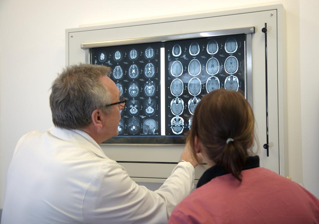
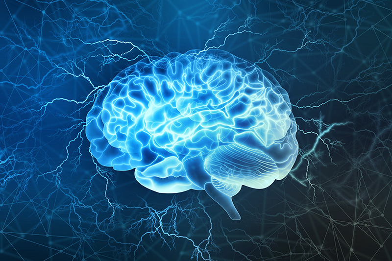
In order to understand the brain, scientists must be able to see the brain — cell by cell, and moment by moment. However, because brains comprise billions of microscopic moving parts, faithfully recording their activity comes with many challenges. In dense mammalian brains, for example, it is difficult to track rapid cellular changes across multiple brain structures — particularly when those structures are located deep within the brain.

A novel microscopy technique, developed by Rockefeller scientists, integrates new and existing approaches to help build a more cohesive picture of the brain. Described in Cell, the technology captures cellular activity across large volumes of neural tissue, with impressive speed and at new depths.

Laser focused

For decades, brain imaging has been plagued by trade-offs. Some techniques produce beautiful images but fail to record neural activity in real time. Others can keep up with the brain’s speed but have poor spatial resolution. And although there are tactics that successfully combine rapidity and image quality, they typically capture only a small number of cells.

“This is in part because the limits that govern these tradeoffs have not been explored or pushed in a systematic and integrated manner,” says Alipasha Vaziri, head of the Laboratory of Neurotechnology and Biophysics.

Hoping to end the era of trade-offs, Vaziri recently endeavored to improve upon a technique known as two-photon (2p) microscopy. It involves the application of a laser that causes bits of brain tissue to fluoresce, or light up; and for many researchers, 2p has long been the gold standard for probing cellular activity in the brain.

Yet, this technique has limitations. Standard 2p microscopy requires point-by-point scanning of a given region, which results in slow imaging. To resolve this issue, Vaziri and his colleagues implemented a novel strategy that permits recording from multiple brain regions in parallel, while carefully controlling the size and shape of each spot recorded.

Another weakness of traditional 2p is that it measures only the surface, or cortex, of the brain, neglecting structures buried deep within the organ, such as the hippocampus, which is involved in storing memories.

“One of the biggest challenges in neuroscience is developing imaging techniques that measure the activity of deep brain regions while maintaining high resolution,” says Vaziri.

Taking up this challenge, he decided to make use of a newer technology: three-photon (3p) microscopy. Whereas 2P doesn’t reach beyond the surface, or cortex, of a mouse brain, 3p penetrates deeper regions. Called hybrid multiplexed sculpted light microscopy, or HyMS, Vaziri’s latest innovation applies 2P and 3P concurrently, allowing researchers to generate a picture of rapid cellular activity across multiple layers of brain tissue.

Deep dive

In addition to its hybrid laser strategy, HyMS also integrates other recent technical and conceptual advancements in the field — a synergistic approach that, Vaziri says, guided the development of the technology. The goal, he says, was to maximize the amount of biological information that could be obtained through multi-photon excitation microscopy while minimizing the heat produced by this method. And when testing their new system, the scientists certainly obtained a lot of information.

HyMS boasts the highest frame rate of available 3p techniques, which means it can capture biological changes at record speed. And whereas previous techniques scanned only a single plane of tissue, this technology can obtain information from the entire tissue sample and allows users to record from as many as 12,000 neurons at once. Another advantage of HyMS is its ability to simultaneously measure activity from brain areas at different depths. Since different layers of the brain constantly exchange signals, says Vaziri, tracking the interplay between these regions is key to understanding how the organ functions.

“Before, people hadn’t even been able to look at the activity of neurons over the entire depth of the cortex, which has multiple layers, all at the same time,” he says. “With this technology you can actually see what the information flow looks like within the cortex, and between cortical and subcortical structures.”

In addition to probing new depths, HyMS allows researchers to record brain activity from animals as they actively engage with their environment. In a recent experiment, for example, the researchers used the technology to record signals from thousands of mouse neurons as an animal walked on a treadmill or listened to sounds. The fact that they were able to obtain good recordings suggests that the technique may be used to monitor large cell populations as animals perform diverse tasks — an application that could help elucidate neural mechanisms underlying various aspects of behavior and cognition.

Further, says Vaziri, techniques like HyMS will be vital to researchers hoping to better understand how brains process information. Neurons in the brain are densely interconnected and information is often represented not by individual cells, but by states of the network.

“To understand the dynamics of a network,” he says, “you need to get accurate measurements of big portions of the brain at a single-neuron level. That’s what we’ve done here.”

New pill can deliver insulin through the stomach

An MIT-led research team has developed a drug capsule that could be used to deliver oral doses of insulin, potentially replacing the injections that people with type 1 diabetes have to give themselves every day.

About the size of a blueberry, the capsule contains a small needle made of compressed insulin, which is injected after the capsule reaches the stomach. In tests in animals, the researchers showed that they could deliver enough insulin to lower blood sugar to levels comparable to those produced by injections given through skin. They also demonstrated that the device can be adapted to deliver other protein drugs.

“We are really hopeful that this new type of capsule could someday help diabetic patients and perhaps anyone who requires therapies that can now only be given by injection or infusion,” says Robert Langer, the David H. Koch Institute Professor, a member of MIT’s Koch Institute for Integrative Cancer Research, and one of the senior authors of the study.

Giovanni Traverso, an assistant professor at Brigham and Women’s Hospital, Harvard Medical School, and a visiting scientist in MIT’s Department of Mechanical Engineering, where he is starting as a faculty member in 2019, is also a senior author of the study. The first author of the paper, which appears in the Feb. 7 issue of Science, is MIT graduate student Alex Abramson. The research team also includes scientists from the pharmaceutical company Novo Nordisk.

Self-orientation

Several years ago, Traverso, Langer, and their colleagues developed a pill coated with many tiny needles that could be used to inject drugs into the lining of the stomach or the small intestine. For the new capsule, the researchers changed the design to have just one needle, allowing them to avoid injecting drugs into the interior of the stomach, where they would be broken down by stomach acids before having any effect.

The tip of the needle is made of nearly 100 percent compressed, freeze-dried insulin, using the same process used to form tablets of medicine. The shaft of the needle, which does not enter the stomach wall, is made from another biodegradable material.

Within the capsule, the needle is attached to a compressed spring that is held in place by a disk made of sugar. When the capsule is swallowed, water in the stomach dissolves the sugar disk, releasing the spring and injecting the needle into the stomach wall.

The stomach wall has no pain receptors, so the researchers believe that patients would not be able to feel the injection. To ensure that the drug is injected into the stomach wall, the researchers designed their system so that no matter how the capsule lands in the stomach, it can orient itself so the needle is in contact with the lining of the stomach.

“As soon as you take it, you want the system to self-right so that you can ensure contact with the tissue,” Traverso says.

The researchers drew their inspiration for the self-orientation feature from a tortoise known as the leopard tortoise. This tortoise, which is found in Africa, has a shell with a high, steep dome, allowing it to right itself if it rolls onto its back. The researchers used computer modeling to come up with a variant of this shape for their capsule, which allows it to reorient itself even in the dynamic environment of the stomach.

“What’s important is that we have the needle in contact with the tissue when it is injected,” Abramson says. “Also, if a person were to move around or the stomach were to growl, the device would not move from its preferred orientation.”

Once the tip of the needle is injected into the stomach wall, the insulin dissolves at a rate that can be controlled by the researchers as the capsule is prepared. In this study, it took about an hour for all of the insulin to be fully released into the bloodstream.

Easier for patients

In tests in pigs, the researchers showed that they could successfully deliver up to 300 micrograms of insulin. More recently, they have been able to increase the dose to 5 milligrams, which is comparable to the amount that a patient with type 1 diabetes would need to inject.

After the capsule releases its contents, it can pass harmlessly through the digestive system. The researchers found no adverse effects from the capsule, which is made from biodegradable polymer and stainless steel components.

Maria José Alonso, a professor of biopharmaceutics and pharmaceutical technology at the University of Santiago de Compostela in Spain, describes the new capsule as a “radically new technology” that could benefit many patients.

“We are not talking about incremental improvements in insulin absorption, which is what most researchers in the field have done so far. This is by far the most realistic and impactful breakthrough technology disclosed until now for oral peptide delivery,” says Alonso, who was not involved in the research.

The MIT team is now continuing to work with Novo Nordisk to further develop the technology and optimize the manufacturing process for the capsules. They believe this type of drug delivery could be useful for any protein drug that normally has to be injected, such as immunosuppressants used to treat rheumatoid arthritis or inflammatory bowel disease. It may also work for nucleic acids such as DNA and RNA.

“Our motivation is to make it easier for patients to take medication, particularly medications that require an injection,” Traverso says. “The classic one is insulin, but there are many others.”

The research was funded by Novo Nordisk, the National Institutes of Health, a National Science Foundation Graduate Research Fellowship, Brigham and Women’s Hospital, a Viking Olaf Bjork Research Scholarship, and the MIT Undergraduate Research Opportunities Program.

Other authors of the paper include Ester Caffarel-Salvador, Minsoo Khang, David Dellal, David Silverstein, Yuan Gao, Morten Revsgaard Frederiksen, Andreas Vegge, Frantisek Hubalek, Jorrit Water, Anders Friderichsen, Johannes Fels, Rikke Kaae Kirk, Cody Cleveland, Joy Collins, Siddartha Tamang, Alison Hayward, Tomas Landh, Stephen Buckley, Niclas Roxhed, and Ulrik Rahbek.

Scientists discover how neuroactive steroids dampen inflammatory signaling in cells

For the first time, scientists discovered how neuroactive steroids naturally found in the brain and bloodstream inhibit the activity of a specific kind of protein called Toll-like receptors (TLR4), which have been known to play a role in inflammation in many organs, including the brain.

This UNC School of Medicine-University of Maryland collaboration, published in Nature Scientific Reports, shows how the neurosteroid allopregnanolone prevents the activation of pro-inflammatory proteins important for gene regulation, as well as the creation of cytokines, which are known to be involved in many different inflammatory conditions. Inflammatory cell signaling in the brain is heightened in many neuropsychiatric conditions, including alcohol use disorder, depression, and posttraumatic stress. It is also seen in sepsis, epilepsy, multiple sclerosis, and Alzheimer’s disease.

“It has been very difficult to treat brain disease that involves inflammation, but allopregnanolone’s inhibition of TLR4 signaling activation in macrophages and the brain provides hope that we can develop better therapies to help millions of people suffering with these conditions,” said senior author A. Leslie Morrow, PhD, the John Andrews Distinguished Professor in the Departments of Psychiatry and Pharmacology at the UNC School of Medicine.

Neuroactive steroids, which are naturally occurring steroids in the brain and elsewhere in the body, have many functions critical for life and health. These steroids decline with aging and are deficient in many neuropsychiatric conditions, such as depression. Morrow and her colleagues have proposed that treatment with these compounds may prevent uncontrolled TLR4 signaling in conditions where this signaling contributes to disease.

Recent studies showed that the neurosteroid compounds pregnenolone and allopregnanolone have therapeutic effects in depression, schizophrenia and PTSD. But until now, scientists didn’t understand how. The UNC-Maryland study suggests that inhibition of inflammatory signaling may contribute to these effects, and inhibition of TLR4 signaling may be a new target for these conditions.

In collaboration with Laure Aurelian, PhD, at the University of Maryland, Morrow and colleagues found that allopregnanolone inhibits TLR4 activation in macrophages, which are found in white blood cells and part of the immune system, including in the brain. In particular, the researchers found that allopregnanolone prevents TLR4 binding to MD2 proteins that work together to produce transcription factors that regulate the genes responsible for inflammatory responses in cells and tissues. Allopregnanolone also tamps down chemokines and cytokines, such as NFkB, HMGB1, MCP-1 and TNF-a, all of which are part of the immune system and involved in many different inflammatory diseases.

Morrow and colleagues found that pregnenolone also inhibited TLR4 signaling in macrophage cells. “Pregnenolone’s effects in the brain were less pronounced,” Morrow said. “But inhibition of peripheral inflammation protects the brain as well because systemic inflammation affects organs throughout the body indirectly.”

Now that scientists have identified this inhibitory mechanism that dampens inflammatory signals responsible for brain inflammation, researchers can create new compounds to fill this particular role of neurosteroids without unwanted side effects. In addition, researchers can now plan clinical studies to determine the best doses, formulations, and modes of administration for different conditions.

David Rubinow, MD, chair of the department of psychiatry at UNC-Chapel Hill, who was not involved in the study, said, “This great example of collaborative and translational research provides physiologic insights with great potential for spawning new, more effective primary and adjunctive treatments for the many individuals suffering from brain disorders characterized by so-called neuroinflammation.”

Scientists design ‘smart’ wound healing technique

New research, published in the journal Advanced Materials, paves the way for “a new generation of materials that actively work with tissues to drive [wound] healing.”

As more and more surgical procedures are performed in the United States, the number of surgical site infections is also on the rise.

Chronic wounds that do not heal — such as those that occur in diabetes — often host a wide range of bacteria in the form of a biofilm.

Such biofilm bacteria are often very resilient to treatment, and antimicrobial resistance only increases the possibility that these wounds become infected.

According to recent estimates, chronic wounds affect approximately 5.7 million people in the U.S. Some chronic wounds can result in amputations, as is the case with diabetic ulcers.

On a global level, researchers approximate that every 30 seconds a chronic, nonhealing diabetic ulcer causes an amputation.

In this context, there is a dire need for innovative, effective wound healing methods. New research shows promise in this regard, as scientists have devised a molecule that helps harness the body’s natural healing powers.

The molecules are called traction force-activated payloads (TrAPs). They are growth factors that help materials such as collagen interact with the body’s tissues more naturally.

Ben Almquist, Ph.D., a lecturer in the department of engineering at Imperial College London in the United Kingdom, led the new research.

TrAP technology and wound healing

Materials such as collagen are often used in wound healing. For instance, collagen sponges can treat burn injuries, and collagen implants can help bones regenerate.

But how does collagen interact with tissue? In so-called scaffold implants, cells move through the collagen structure, pulling the scaffold along with them. This triggers healing proteins, such as growth factors, that help the tissue regenerate.

In the new study, Almquist and the team engineered TrAP molecules to recreate this natural process. The scientists “folded” DNA strands into aptamers, which are three-dimensional shapes that bind to proteins.

Then, they designed a “handle” for cells to grip. They attached cells to one end of the handle and a collagen scaffold to the other end.

Lab tests revealed that the cells dragged the TrAPs along as they moved through the collagen implants. In turn, this activated growth proteins that triggered the healing process within the tissue.

The scientists explain that this technique recreates healing processes that exist throughout the natural world. “Using cell movement to activate healing is found in creatures ranging from sea sponges to humans,” says Almquist.

“Our approach mimics them and actively works with the different varieties of cells that arrive in our damaged tissue over time to promote healing,” he adds.

A ‘new generation’ of healing materials

The research also revealed that tweaking the cellular handle changes the type of cells that can attach and hold on to the TrAPs.

In turn, this enables TrAPs to release personalized regenerative proteins based on the cells that have attached to the handle.

This adaptability to different types of cells means that the technique can be applied to various types of wounds — ranging from bone fractures to scar tissue injuries caused by heart attacks and from nerve damage to diabetic ulcers.

Finally, aptamers are already approved as drugs for human clinical use, which could mean that the TrAP technique may become widely available sooner rather than later.

“The TrAP technology provides a flexible method to create materials that actively communicate with the wound and provide key instructions when and where they are needed,” explains Almquist.

“This sort of intelligent, dynamic healing is useful during every phase of the healing process, has the potential to increase the body’s chance to recover, and has far-reaching uses on many different types of wounds,” he adds.

The researcher concludes, “[t]his technology has the potential to serve as a conductor of wound repair, orchestrating different cells over time to work together to heal damaged tissues.”

High-tech epilepsy warning device could save lives

Scientists have developed a high-tech bracelet called the Nightwatch, which detects 85 percent of all severe nighttime epilepsy seizures — a much higher percentage than similar devices on the market today.
Brain scans check
A new type of wearable technology may help prevent epilepsy-related deaths.

Smartwatches are gaining popularity, and they often help users monitor their health in different ways, such as by recording sleeping habits or heart rates.

The researchers, who published their results in the journal Neurology, believe that this bracelet could be a vital tool for people with epilepsy.

Sudden unexpected death in epilepsy (SUDEP) is the leading cause of death in those with the condition. The risks of death are even higher in people who have therapy-resistant epilepsy and an intellectual disability.

The research team, based in the Netherlands, tested the Nightwatch with 28 intellectually disabled participants who have epilepsy.

Testing the Nightwatch

Each participant wore the bracelet for an average of 65 nights, and the Nightwatch was set to sound an alarm if the person had a severe seizure. The participants were filmed to determine if there were any false alarms or seizures that the device failed to catch.

The Nightwatch works by recognizing two specific characteristics of severe epileptic seizures — a very rapid heartbeat and rhythmic, jolting movements. When these are detected, the device will immediately send an alert to the person’s caregiver.

Overall, the device recognized 85 percent of all severe attacks and 96 percent of those that were the most severe.

Both scores are far higher than those of similar devices. The current standard method of detection is a bed sensor that reacts to vibrations caused by rhythmic jerks.

The researchers also tested this sensor, for comparison, and it only detected severe seizures 21 percent of the time.

When the data were tabulated, the Nightwatch had missed a serious attack once out of every 25 nights per patient, which is far less often than the bed sensor — this had missed a serious seizure once out of every 4 nights per patient.

Epilepsy and the risk of death

While epilepsy involves seizures, not everyone who has experienced a seizure has the condition.

Instead, epilepsy is defined as a chronic, ongoing disorder hallmarked by recurrent and unprovoked seizures.

There are a number of types of epilepsy, defined by several factors.

When making a diagnosis, a neurologist will take into account what types of seizures the person experiences, how old they were when the seizures began, what part of the brain is involved, and what patterns can be detected, among other considerations.

Epilepsy is not always fatal, but those with this neurological disorder are at risk, in some cases. The leading cause of epilepsy-related death, as mentioned above, is SUDEP.

Following SUDEP, the body is often found in bed. In only a third of cases, there is evidence that the person experienced a seizure close to the time of death. Also, the body is frequently found facedown, which leads researchers to consider that suffocation may be involved.

There are a number of risk factors for SUDEP, including being aged 20–40, experiencing seizures at night, and having epilepsy that began during childhood.

In addition, those who have poorly controlled epilepsy are at much greater risk than those who do not, including patients that do not take their medication as scheduled. Also at increased risk are those with therapy-resistant epilepsy.

The Nightwatch may be a valuable tool for people at risk of SUDEP, and it could make a resounding difference for epilepsy patients, their caregivers, and their families.

The research leader, professor and neurologist Dr. Johan Arends, says that the device may reduce the number of SUDEP incidences by two-thirds.

However, he notes that this figure will depend on how quickly carers respond to the sounded alerts. If the device finds its way around the globe, it may help save thousands of lives.

Half a million tests and many mosquitoes later, new buzz about a malaria prevention drug

Most malaria drugs are designed to reduce symptoms after infection. They work by blocking replication of the disease-causing parasites in human blood, but they don’t prevent infection or transmission via mosquitoes. What’s worse, the malaria parasite is developing resistance to existing drugs.

“In many ways, the search for new malaria drugs has been a search for something akin to aspirin — it makes you feel better but doesn’t necessarily go after the root of the problem,” said Elizabeth Winzeler, PhD, professor of pharmacology and drug discovery at University of California San Diego School of Medicine.

In a study publishing December 7 in Science, Winzeler and her team took a different approach: targeting the malaria parasite at an earlier stage in its lifecycle, when it initially infects the human liver, rather than waiting until the parasite is replicating in blood and making a person ill.

The team spent two years extracting malaria parasites from hundreds of thousands of mosquitoes and using robotic technology to systematically test more than 500,000 chemical compounds for their ability to shut down the malaria parasite at the liver stage. After further testing, they narrowed the list to 631 promising compounds that could form the basis for new malaria prevention drugs.

To help speed this effort, the researchers made the findings open source, meaning the data are freely shared with the scientific community.

“It’s our hope that, since we’re not patenting these compounds, many other researchers around the world will take this information and use it in their own labs and countries to drive antimalarial drug development forward,” Winzeler said.

Most cases of malaria are caused by the mosquito-borne parasites Plasmodium falciparum or Plasmodium vivax. The parasites’ lifecycle begins when an infected mosquito transmits sporozoites into a person while taking a blood meal. A few of these sporozoites may establish an infection in the liver. After replicating there, the parasites burst out and infect red blood cells. That’s when the person begins to experience malarial symptoms, such as fever, chills and headaches. That’s also when the parasite can be sucked up by a new mosquito and transmitted to another person.

For safety’s sake, Winzeler and team used a related parasite called Plasmodium berghei in the study, which can only infect mice. Their collaborators in New York infected mosquitos with these parasites and every Tuesday, Winzeler’s team would receive a big orange box of mosquitoes by FedEx. On Tuesday afternoons, they would extract the sporozoites, transfer them to plates containing 1,536 tiny divots, or wells, and then carry the plates over to the drug screening facilities at Skaggs School of Pharmacy and Pharmaceutical Sciences at UC San Diego or across the street to the Genomics Institute of the Novartis Research Foundation.

“In a good week, we’d be able to test 20,000 compounds,” Winzeler said, “but of course many of the mosquitoes we received would be dried out, frozen or covered in fungus.”

These sporozoites were engineered to produce luciferase, the same enzyme that fireflies use to produce their telltale glow. Then, in the drug screening facilities, researchers used robotic technology and sound waves to add minute amounts of each chemical compound, one compound per sporozoite-containing well.

The researchers looked for the compounds that switch the glow “off,” meaning they had killed the parasites or blocked their ability to replicate. They took those compounds, confirmed their potency and weeded out the ones toxic to liver cells. They also tested the compounds for their ability to inhibit or kill other Plasmodium species and parasites at other lifecycle stages.

That left them with 631 promising chemical compounds — chemical starting points for the development of new drugs to block a malaria infection before symptoms begin, and prevent their transmission to the blood, mosquitoes and other people. Winzeler said she was surprised to find that many compounds (58) block the parasite’s electron transport chain, an energy-generating process in its mitochondria.

The team will next take a closer look at the 631 promising drug candidates to determine how many work against the liver stage of the Plasmodium species that affect humans. Winzeler and members of the Bill and Melinda Gates Foundation Malaria Drug Accelerator (MalDA), an international consortium focused on speeding drug development, are collaborating to unravel the mechanism by which many of the compounds work against the malaria parasite.

The team and others will also continue the work necessary to develop the compounds into drugs that are safe for human consumption and effective at preventing liver-stage parasites from replicating and bursting out into the bloodstream. The ideal new drug would also be affordable and practical for administration in parts of the world without refrigeration or an abundance of health care providers.

“It’s difficult for many people to consistently sleep under mosquito nets or take a daily pill,” Winzeler said. “We’ve developed many other options for things like birth control. Why not malaria? The malaria research community has always been particularly collaborative and willing to share data and resources, and that makes me optimistic that we’ll soon get there too.”

According to a new report from the World Health Organization (WHO), malaria cases are on the rise, particularly in 13 countries, including Madagascar, Nigeria and the Democratic Republic of the Congo. There were 219 million cases of malaria in 2017, compared to 217 million the previous year. In 2017, approximately 435,000 people died of malaria.

Mutations boost immunity: Toward a cancer vaccine

Despite significant advances in cancer research, the disease continues to exact a devastating toll. Because cancer is a disease of the body’s own cells, which mutate and develop under evolutionary pressure, conventional treatments like chemotherapy and radiation often leave behind a residue of resistant cells that go on to expand and wreak havoc.

The best weapon against this implacable foe would be prevention, though to date, this has been an elusive goal.

In a new study, Stephen Albert Johnston and his colleagues describe a method for pinpointing tumor-specific factors in blood that can elicit a protective immune response in the body and may one day be harnessed to produce an effective vaccine against the disease.

The new study outlines a means for rapidly identifying peptides produced by tumor-associated mutations, then screening these peptides to find those exhibiting a strong immune response.

A new vision

The work is part of a sea change in the field of oncology, where increasingly, the body’s immune system is induced to attack the disease. Immunotherapies have already shown startling effectiveness against certain previously intractable cancers and a pair of scientists were awarded this year’s Nobel Prize for their research into immune mechanisms known as checkpoint inhibitors.

The technique described in the new study relies on libraries of peptides printed on slides known as peptide arrays. When such arrays are exposed to cancer-linked antigens in samples of patient blood, specific peptides bind with antibodies, suggesting they are recognized by the immune system and may be used in a vaccine against that cancer.

Results of the study indicate that tumor-associated peptide mutations not only bind with immune antibodies, but can effectively provide cancer protection, (at least in animal models of the disease). The peptides generating a strong immune response could be incorporated into a vaccine or alternatively, used in conjunction with other forms of immunotherapy to treat existing cancers.

Johnston and his colleagues used peptide arrays to screen for tumor-linked peptides in blood samples from dogs, examining responses to 9 different forms of cancer. The antigens showing the greatest immune response in the array were then evaluated for their protective effect against two forms of cancer, in a mouse model.

The study confirmed that some of the peptides exhibiting a strong antibody response on the peptide arrays offered protection from cancer in mice, while non-immunogenic peptides did not.

“Our system has the advantages of not requiring tumor tissue to DNA sequence and not having to guess whether a mutation elicits an immune response,” Johnston says.

Johnston directs the Biodesign Institute Center for Innovations in Medicine. The new study appears in the journal Scientific Reports.

Hidden in plain sight

When viruses, bacteria or other pathogens attack the body, they often carry particular molecular signatures not present in normal cells. The immune system can recognize these foreign signatures, mounting a defense against the disease-causing invader.

Cancer is different. Because cancer is a disease involving the body’s own native cells, most telltale signs of an alien presence, capable of triggering the immune system, are lacking.

Fortunately, the body is not entirely defenseless against cancer. Certain signposts of illness produced by cancerous tumors can indeed provoke an immune response. Particular mutated peptides can act to alert the immune system, once they have been expressed, processed and presented on the cell surface, allowing the immune system warriors — the T cells — to recognize and attack the cancer.

Identifying and harnessing these factors — known as neoantigens — is the focus of the new study.

But while cancer produces a variety of mutations, whose traces may be registered by the immune system, Johnston notes that not all mutations are created equal. A specific form — known as frameshift mutations — have been shown to be more effective stimulators of immune response. They have been difficult to isolate and identify, until now.

If tumor-specific frameshift mutations can be recognized and applied in cancer therapy, the results are potentially dramatic, because T cells specific to cancer neoantigens can aggressively attack malignant cells without harming normal tissue.

Shifting frames of reference

Most efforts toward a cancer vaccine have focused on so-called point mutations. Such mutations occur when a single DNA nucleotide letter is replaced with a different nucleotide. For example, an original sequence of ACCTACA could mutate to form a sequence reading ACCTATA.

Point mutations therefore leave the sequence length unchanged, altering only the content of the DNA and resulting RNA transcripts. By contrast, frameshift mutations occur when sequence letters are inserted or deleted. (INDELS is the term for these insertion-deletion mutations.)

Currently, use of point mutations for experimental cancer vaccines have been largely based on algorithms that make predictions about which neoantigens will yield an effective immune response, which can only be tested for effectiveness once the vaccine has been manufactured. The process, which is estimated to take 1-3 months, is cumbersome, very expensive and inaccurate. Use of frameshift peptide arrays could provide immediate information on peptide vaccine candidates and assess their immune reactivity before the formulation of vaccines.

In addition to indels, frameshift mutations can occur through a process known as exon mis-splicing. Exon splicing occurs prior to translation from RNA to protein. Here, nucleotide sequences known as introns, which do not code for proteins, are cut from sequences and ends of the remaining coding regions, known as exons, are fused. This process can mis-splice — either omitting part of the exon or including part of the unwanted intron sequence. Like indel mutations, exon mis-splicing is a rich source of immunogenic mutations, explored in the current research.

The search

The new study describes a means of ferreting out tumor-specific peptides resulting from frame shift mutations by preparing peptide arrays containing libraries of frameshift peptides to probe for cancer-specific antibodies to them in dogs, then testing the capacity of the resulting antigens to protect against cancer in a mouse model.

Dogs are subject to a variety of cancers that also plague humans, making them attractive subjects for such a study. Johnston plans to explore both therapeutic and prophylactic vaccines in dogs in parallel to human trials.

As the authors note, there are a finite number of possible peptides displaying frameshift mutations, so it is possible to construct arrays capable of interrogating the entire sequence space of these mutations, eventually establishing the most immunogenic candidates. A group of 10-20 such frameshift peptides could be used for an anti-cancer vaccine.

In the present study, 830 peptides from 377 predicted frameshift antigens were synthesized and affixed to array slides. 116 samples of blood serum from 26 dog breeds, representing 9 types of dog cancer (carcinoma, fibrosarcoma, hemangiosarcoma, lymphoma, mast cell tumor, osteosarcoma, histiocytic sarcoma, synovial cell sarcoma and malignant histiocytosis) were screened on the dog frameshift peptide array. 52 age-matched, blood samples from healthy dogs were used as control. (Each frameshift antigen was represented with 1-4, frameshift peptides, 17 nucleotides in length on the array.)

Subsequent testing of the frameshift peptides demonstrated that reactive peptides provided T cell protection from melanoma and breast cancer in mice, whereas non-reactive peptides offered no such protection. Intriguingly, this tumor protection directly correlated to the degree of antibody response to frameshift peptides seen in the array results.

The research paves the way for the development of potent new weapons against cancer, leveraging the body’s own immune defenses to stop this leading killer in its tracks.

Implants ‘made of your own cells’ could end back pain

Back and neck pain are often the result of the progressive damage of the discs that separate the spinal vertebrae. Thanks to new multidisciplinary research, we may soon have a better solution to this problem: bioengineered discs grown out of a person’s own cells.

Intervertebral disc degeneration is a common problem that affects a large segment of the population.

Typically, healthy intervertebral discs function by absorbing stress placed on the spine as we move and adjust our posture in a similar way to a car suspension.

If those discs wear out, it can cause pain in various areas of a person’s back or neck.

So far, treatments for intervertebral disc degeneration include spinal fusion surgery and replacing the damaged discs with artificial ones.

However, these approaches bring limited benefits because they cannot restore full function of the intervertebral discs they replace.

Now, a multidisciplinary research team from the University of Pennsylvania’s Perelman School of Medicine, School of Engineering and Applied Science, and School of Veterinary Medicine is aiming to solve this issue by developing bioengineered intervertebral discs made out of an individual’s own stem cells.

Stem cells are undifferentiated cells that have the potential to “transform” into any specialized cells. That is why they have become the focus of multiple medical research studies, including the current one.

The researchers at the University of Pennsylvania have been working for the past 15 years on bioengineered disc models — first in laboratory studies, then in small animal studies, and most recently in large animal studies.

“This is a major step: to grow such a large disc in the lab, to get it into the disc space, and then to have it to start integrating with the surrounding native tissue. That’s very promising,” says Prof. Robert L. Mauck, co-senior author of the current study.

“The current standard of care does not actually restore the disc, so our hope with this engineered device is to replace it in a biological, functional way and regain full range of motion,” he adds.

Studies in animal successful so far
Previously, the researchers tested the new discs — called “disc-like angle ply structures” (DAPS) — in rat tails for 5 weeks.

In the new study, whose results appear in the journal Science Translational Medicine, the team developed the engineered discs even further. They then tested the new model — called ” endplate-modified DAPS” (eDAPS) — in rats again, but this time for up to 20 weeks.

The new structure of the bioengineered disc allows it to retain its shape better, and integrate more easily with the surrounding tissue.

Following several tests — MRI scans and several in-depth tissue and mechanical analyses — the researchers found that, in the rat model, eDAPS effectively restored original disc structure and function.

This initial success motivated the research team to study eDAPS in goats, and they implanted the device into the cervical spines of some of the animals. The scientists chose to work with goats because, as they explain, the cervical spinal discs of goats have similar dimensions to those of humans.

Moreover, goats have semi-upright stature, allowing the researchers to bring their study one step closer to human trials.

‘A very good reason to be optimistic’
The researchers’ tests on goats were also successful. They noticed that the eDAPS integrated well with the surrounding tissue, and the mechanic function of the discs at least matched, if not surpassed, that of the original cervical discs of the goats.

“I think it’s really exciting that we have come this far, from the rat tail all the way up to human-sized implants,” says Dr. Harvey E. Smith, co-senior author of the study.

“When you look at the success in the literature from mechanical devices, I think there is a very good reason to be optimistic that we could reach that same success, if not exceed it with the engineered discs.” – Dr. Harvey E. Smith

The researchers say that the next step will include conducting further, more extensive trials in goats, which will allow the scientists to understand better how well eDAPS works.

Moreover, the research team plans to test out eDAPS in models of human intervertebral disc degeneration, thus hopefully getting one step closer to clinical trials.

“There is a lot of desirability to implant a biological device that is made of your own cells,” notes Dr. Smith, adding that, “Using a true tissue-engineered motion-preserving replacement device in arthroplasty of this nature is not something we have yet done in orthopaedics.”

“I think it would be a paradigm shift for how we really treat these spinal diseases and how we approach motion sparing reconstruction of joints,” he continues.

New drug options, risk factors added to U.S. heart guidelines

The recommendations from the American Heart Association and the American College of Cardiology, last issued in 2013, acknowledge recent research showing the benefit of very low levels of “bad” LDL cholesterol, which contributes to fatty plaque buildup and narrowing of arteries.

The medical groups, which announced the guidelines on Saturday at the AHA’s annual meeting in Chicago, still emphasize a healthy diet and exercise as the first line of defense against heart disease, the No. 1 killer in the country.

When cholesterol is not controlled by lifestyle efforts, patients are typically prescribed statin drugs, available as low cost generic pills that have long been proven to safely and effectively lower LDL levels and heart disease risk.

For people who have had a heart attack or stroke, are at high risk for another and whose cholesterol levels are not adequately lowered by statins, the guidelines now recommend adding newer cholesterol drugs.

Higher risk patients are advised to first try statins in combination with ezetimibe, the generic version of Zetia, which lowers cholesterol by limiting its absorption from the intestine. If that does not work, the guidelines call for newer injected medications known as PCSK9 inhibitors, specifically for people who are at very high risk or who have a genetic condition that causes very high cholesterol levels.

Two PCSK9 drugs – Amgen Inc’s (AMGN.O) Repatha and Praluent from partners Regeneron Pharmaceuticals Inc (REGN.O) and Sanofi SA (SASY.PA) – were launched in 2015 at U.S. prices of over $14,000 a year.

Both have been shown to dramatically lower cholesterol levels, but uptake as been slow as the medical community and health insurers questioned their cost effectiveness.

To help spur usage, Amgen last month cut Repatha’s list price by 60 percent to $5,850 and Praluent’s annual net price was cut earlier this year to between $4,500 and $6,600.

The new guidelines are fairly “conservative” in recommending that the newer drugs be used only after other options, said Dr. Francisco Lopez-Jimenez, a cardiologist at the Mayo Clinic in Rochester, Minnesota, adding “I think that was the right approach.”

The guidelines continue to include a calculator introduced in 2013 to identify a patient’s 10-year risk for cardiovascular disease. In addition to traditional risk factors such as smoking and high blood pressure, doctors are now urged to discuss family history and ethnicity as well as health conditions such as chronic kidney disease and premature menopause.

They are also advised to test for cholesterol levels in children as young as two with a family history of heart disease or high cholesterol, while other children should have an initial test between the ages of nine and 11.

Coronary artery calcium measurements are advised for people whose risk level is not clear.

The guidelines update has more specific recommendations for certain age and ethnic groups, as well as for people with diabetes.

Long-lasting HIV injection is a step closer after second GSK study

LONDON (Reuters) – A once-monthly injection to control HIV proved as effective as daily pills in a second study by GlaxoSmithKline, paving the way for a new regimen that could be simpler for some patients to be filed with regulators.

The experimental two-drug injection of cabotegravir and rilpivirine was shown to suppress the HIV virus in a cohort of adults who had not been on a long-established daily three-drug oral regimen, GSK’s majority-owned HIV unit ViiV Healthcare said.

In the study, adults with HIV were first put on a 20-week three-drug program of daily tablets to suppress the virus before being switched to monthly injections. After 48 weeks, the injections maintained a similar rate of suppression as the pills, it said.

The results support an earlier major study, which involved adults who had been using a three-drug oral regimen to control the virus.

ViiV Healthcare’s chief medical officer John C. Pottage said the trial provided further evidence that a long-acting injection could offer an alternative to daily, oral therapy for people who had previously achieved viral suppression.

“This innovative dosing regimen could transform HIV therapy by reducing the number of days a person receives treatment from 365 to 12,” he said.

“Work on new methods of HIV treatment, including long-acting injectable therapies, supports our goal of making HIV a smaller part of the lives of people living with HIV.”

ViiV Healthcare, which is also owned by Pfizer and Shionogi, hopes that its work on developing two-drug therapies will help it compete against Gilead Sciences, the U.S. drugmaker that dominates the $26 billion-a-year HIV market.

The company said detailed results of the study would be presented at an upcoming scientific meeting.