Scientists design ‘smart’ wound healing technique

New research, published in the journal Advanced Materials, paves the way for “a new generation of materials that actively work with tissues to drive [wound] healing.”

As more and more surgical procedures are performed in the United States, the number of surgical site infections is also on the rise.

Chronic wounds that do not heal — such as those that occur in diabetes — often host a wide range of bacteria in the form of a biofilm.

Such biofilm bacteria are often very resilient to treatment, and antimicrobial resistance only increases the possibility that these wounds become infected.

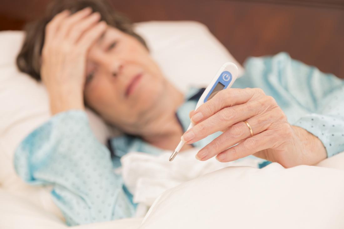
According to recent estimates, chronic wounds affect approximately 5.7 million people in the U.S. Some chronic wounds can result in amputations, as is the case with diabetic ulcers.

On a global level, researchers approximate that every 30 seconds a chronic, nonhealing diabetic ulcer causes an amputation.

In this context, there is a dire need for innovative, effective wound healing methods. New research shows promise in this regard, as scientists have devised a molecule that helps harness the body’s natural healing powers.

The molecules are called traction force-activated payloads (TrAPs). They are growth factors that help materials such as collagen interact with the body’s tissues more naturally.

Ben Almquist, Ph.D., a lecturer in the department of engineering at Imperial College London in the United Kingdom, led the new research.

TrAP technology and wound healing

Materials such as collagen are often used in wound healing. For instance, collagen sponges can treat burn injuries, and collagen implants can help bones regenerate.

But how does collagen interact with tissue? In so-called scaffold implants, cells move through the collagen structure, pulling the scaffold along with them. This triggers healing proteins, such as growth factors, that help the tissue regenerate.

In the new study, Almquist and the team engineered TrAP molecules to recreate this natural process. The scientists “folded” DNA strands into aptamers, which are three-dimensional shapes that bind to proteins.

Then, they designed a “handle” for cells to grip. They attached cells to one end of the handle and a collagen scaffold to the other end.

Lab tests revealed that the cells dragged the TrAPs along as they moved through the collagen implants. In turn, this activated growth proteins that triggered the healing process within the tissue.

The scientists explain that this technique recreates healing processes that exist throughout the natural world. “Using cell movement to activate healing is found in creatures ranging from sea sponges to humans,” says Almquist.

“Our approach mimics them and actively works with the different varieties of cells that arrive in our damaged tissue over time to promote healing,” he adds.

A ‘new generation’ of healing materials

The research also revealed that tweaking the cellular handle changes the type of cells that can attach and hold on to the TrAPs.

In turn, this enables TrAPs to release personalized regenerative proteins based on the cells that have attached to the handle.

This adaptability to different types of cells means that the technique can be applied to various types of wounds — ranging from bone fractures to scar tissue injuries caused by heart attacks and from nerve damage to diabetic ulcers.

Finally, aptamers are already approved as drugs for human clinical use, which could mean that the TrAP technique may become widely available sooner rather than later.

“The TrAP technology provides a flexible method to create materials that actively communicate with the wound and provide key instructions when and where they are needed,” explains Almquist.

“This sort of intelligent, dynamic healing is useful during every phase of the healing process, has the potential to increase the body’s chance to recover, and has far-reaching uses on many different types of wounds,” he adds.

The researcher concludes, “[t]his technology has the potential to serve as a conductor of wound repair, orchestrating different cells over time to work together to heal damaged tissues.”

Pediatric leukemia ‘super drug’ could be developed in the coming years

Northwestern Medicine scientists have discovered two successful therapies that slowed the progression of pediatric leukemia in mice, according to three studies published over the last two years in the journal Cell, and the final paper published Dec. 20 in Genes & Development.

When a key protein responsible for leukemia, MLL, is stabilized, it slows the progression of the leukemia, the most recent study found. The next step will be to combine the treatments from the past two years of research into a pediatric leukemia “super drug” to test on humans in a clinical trial.

The survival rate is only 30 percent for children diagnosed with MLL-translocation leukemia, a cancer that affects the blood and bone marrow. Patients with leukemia have a very low percentage of red blood cells, making them anemic, and have approximately 80 times more white blood cells than people without cancer.

“These white blood cells infiltrate many of the tissues and organs of the affected individuals and is a major cause of death in leukemia patients,” said senior author Ali Shilatifard, the Robert Francis Furchgott Professor of Biochemistry and Molecular Genetics and Pediatrics, the chairman of biochemistry and molecular genetics and the director of Northwestern’s Simpson Querrey Center for Epigenetics. “This is a monster cancer that we’ve been dealing with for many years in children.”

There are several types of leukemia. This research focused on the two most common found in infants through teenagers: acute myeloid leukemia (AML) and acute lymphocytic leukemia (ALL).

For the past 25 years, Shilatifard’s laboratory has been studying the molecular function of MLL within its complex known as COMPASS (Complex Proteins Associated with Set1). Most recently, it was demonstrated that COMPASS components are one of the most frequently identified mutations in cancer. The next step of this work will be to bring the drug to a clinical trial setting, which Shilatifard said he hopes will happen in the next three to five years.

“I’ve been working on this translocation for more than two decades, and we’re finally at the point where in five to 10 years, we can get a drug in kids that can be effective,” Shilatifard said. “If we can bring that survival rate up to 85 percent, that’s a major accomplishment.”

Earlier work from Shilatifard’s laboratory published in Cell in 2018 identified compounds that could slow cancer growth by interrupting a gene transcription process known as “Super Elongation Complex” (SEC). It was the first compound in its class to do this.

This MLL stabilization process discovered in the most recent paper could potentially work in cancers with solid tumors, such as breast or prostate cancer, said first author Zibo Zhao, a postdoctoral research fellow in Shilatifard’s lab.

“This opens up a new therapeutic approach not only for leukemia, which is so important for the many children who are diagnosed with this terrible cancer, but also for other types of cancers that plague the population,” Zhao said.

“The publication of these four papers and the possibility of a future human clinical trial could not have happened if it weren’t for the cross-disciplinary collaboration at Northwestern,” Shilatifard said.

Iron-Rich Foods

Spinach may not give you superhuman strength to fight off villains like Popeye’s nemesis Bluto, but this leafy green and other foods containing iron can help you fight a different type of enemy — iron-deficiency anemia.

Iron-deficiency anemia, the most common form of anemia, is a decrease in the number of red blood cells caused by too little iron. Without sufficient iron, your body can’t produce enough hemoglobin, a substance in red blood cells that makes it possible for them to carry oxygen to the body’s tissues. As a result, you may feel weak, tired, and irritable.

About 20% of women, 50% of pregnant women, and 3% of men do not have enough iron in their body. The solution, in many cases, is to consume more foods high in iron.

How Your Body Uses Iron in Food

When you eat food with iron, iron is absorbed into your body mainly through the upper part of your small intestine.

There are two forms of dietary iron: heme and nonheme. Heme iron is derived from hemoglobin. It is found in animal foods that originally contained hemoglobin, such as red meats, fish, and poultry (meat, poultry, and seafood contain both heme and non-heme iron). Your body absorbs the most iron from heme sources. Most nonheme iron is from plant sources.

Iron-Rich Foods

Very good sources of heme iron, with 3.5 milligrams or more per serving, include:
Continue Reading Below
you might like

  • 3 ounces of beef or chicken liver
  • 3 ounces of clams or mussels
  • 3 ounces of oysters

Good sources of heme iron, with 2.1 milligrams or more per serving, include:

  • 3 ounces of cooked beef
  • 3 ounces of canned sardines, canned in oil

Other sources of heme iron, with 0.6 milligrams or more per serving, include:

  • 3 ounces of chicken
  • 3 ounces of cooked turkey
  • 3 ounces of ham
  • 3 ounces of veal

Other sources of heme iron, with 0.3 milligrams or more per serving, include:

  • 3 ounces of halibut, haddock, perch, salmon, or tuna

Iron in plant foods such as lentils, beans, and spinach is nonheme iron. This is the form of iron added to iron-enriched and iron-fortified foods. Our bodies are less efficient at absorbing nonheme iron, but most dietary iron is nonheme iron.

Read Full Article

Left brain vs. right brain: How does one dominate?

The two hemispheres of our brain — left and right — specialize in different tasks. A recent study asks how this occurs and reaches a surprising conclusion.

Hemispheric dominance, also known as lateralization of brain function, describes the tendency for either the left or the right side of the brain to carry out specific brain activities.

Even though both sides of the brain are almost identical, one hemisphere primarily carries out some functions over others.

For instance, the left hemisphere houses brain regions linked to speech (or the right hemisphere in left-handed people).

Previously, scientists thought humans were the only creatures to exhibit this phenomenon. However, recent research has found lateralized brain function throughout the animal kingdom — from insects, such as honeybees, to aquatic mammals, including killer whales.

The corpus callosum — a thick tract of nerve cells, known as commissural fibers — connects the two hemispheres. Exactly how dominance is produced remains uncertain.

Recently, researchers from Ruhr-Universität Bochum in Germany set out to investigate this question. They chose to study the visual system of pigeons, and they have published their findings in the journal Cell Reports.

Bird brains and old ideas

Previously, scientists have theorized that one side of the brain simply inhibits the other, allowing it to take dominance.

Co-lead author Prof. Onur Güntürkün explains that “[i]n the past, it had been assumed that the dominant hemisphere transmits inhibitory signals to the other hemisphere via the commissures, thus suppressing specific functions in that region.”

In effect, the dominant hemisphere is thought to overpower its neighbor. However, scientists have also noted that excitatory messages run both ways, so there must be more to this interaction.

The researchers decided to use a pigeon model because other studies have described hemispheric dominance in this species in some detail over recent years.

For instance, in pigeon brains, the left hemisphere takes the lead when it comes to visual processing of patterns and colors. Conversely, the right brain more often deals with social or emotionally charged stimuli.

The scientists trained the birds to perform a color differentiation task. In particular, this challenge involves part of the brain that uses visual information to guide motor activity. In this type of task, the left side of the brain is dominant.

To understand how cross-talk between hemispheres influences dominance, Prof. Güntürkün and co-author Dr. Qian Xiao intermittently switched off some of the neurons that run between the two sides of the brain.

Interfering in cross-talk

After blocking specific neurons running from one side, they would observe the activity of the neurons that usually receive their input on the opposite side. In this way, they could pick apart the way in which the dominant hemisphere exerts its control.

The researchers showed that, rather than merely inhibiting the right side of the brain during this task, the left brain could delay the response of the right brain, so preventing it from getting involved.

As Prof. Güntürkün explains, “The right hemisphere simply acts too late to control the response.”

Rather than merely inhibiting the response, the right brain still operates, but its signals are too late to the party to make a difference to the bird’s behavior.

“These results show that hemispheric dominance is based on a sophisticated mechanism. It does not hinge on one general inhibitory or excitatory influence; rather it is caused by minute temporal delays in the activity of nerve cells in the other hemisphere.”

Prof. Onur Güntürkün

The findings provide an entirely new way to look at hemispheric dominance. Research is bound to continue into this rather peculiar phenomenon that evolution has lovingly conserved throughout many branches of life.

However, it is likely to be quite some time before we understand why dividing tasks between the hemispheres is so evolutionarily advantageous.

People who understand the benefits of exercise may spend more time being active

(Reuters Health) – The more people understand about the benefits of exercise, the more time they’re likely to spend being physically active, an Australian study suggests.

Researchers at Central Queensland University surveyed 615 people to explore their knowledge of the benefits of physical activity and the risks of inactivity. The survey also included questions to measure time spent in walking activity, moderate intensity activity (e.g., gentle swimming) and vigorous intensity activity (e.g., cycling).

Writing in PLoS One, senior author Stephanie Schoeppe and colleagues say regular physical activity “reduces the risk of all-cause mortality by 30 percent, reduces the risk of developing major chronic diseases such as cardiovascular disease by 35 percent, type 2 diabetes by 42 percent, (and) colon cancer by 30 percent.”

Furthermore, they write, “Regular physical activity also “increases life expectancy . . . (and) improves general physical health and well-being.”

And indeed, nearly all of those surveyed agreed physical activity is good for health.

On average, however, participants could identify only 14 out of 22 diseases associated with physical inactivity.

And the majority were unable to accurately estimate the increased risk of disease resulting from inactivity.

More than half did not know how much physical activity is recommended for health benefits. (Similar to U.S. guidelines, guidelines in Australia advise adults ages 18 to 64 to engage in at least 150 minutes of moderate intensity or 75 minutes of vigorous intensity physical activity per week.)

Participants were significantly more active when they correctly identified more diseases associated with physical inactivity, the researchers found.

Given the knowledge gaps revealed by their survey, they say, health promotion initiatives should aim to raise awareness of the types of diseases associated with inactivity.

One limitation the authors flagged was that about three quarters of the survey’s respondents were women, so it is unclear whether the results will be applicable to men. Further, the study participants were not representative of the Australian population as a whole.

“A large proportion of Australian adults are insufficiently active,” Schoeppe noted in an email to Reuters Health.

“For those people,” she said, “the messages, ‘any physical activity is better than none’ and ‘doing more physical activity improves health’ are useful.”

Ada Tang, a physical therapist and associate professor at McMaster University, who was not involved in the study, told Reuters Health by email that even if people appreciate the potential risks associated with being physically inactive, they wouldn’t necessarily feel any immediate urgency to act on it.

“The risk of developing heart disease might seem too far in the distant future to drive a person to change their behavior,” she said.

Tang believes initiatives must go beyond improved public relations efforts.

“It is important for us to find ways for people to participate in physical activity more often and more easily,” she said.

“When physical activity becomes more of a habit, it’s not seen as something that is an added burden to their very busy lives.”

High-tech epilepsy warning device could save lives

Scientists have developed a high-tech bracelet called the Nightwatch, which detects 85 percent of all severe nighttime epilepsy seizures — a much higher percentage than similar devices on the market today.
Brain scans check
A new type of wearable technology may help prevent epilepsy-related deaths.

Smartwatches are gaining popularity, and they often help users monitor their health in different ways, such as by recording sleeping habits or heart rates.

The researchers, who published their results in the journal Neurology, believe that this bracelet could be a vital tool for people with epilepsy.

Sudden unexpected death in epilepsy (SUDEP) is the leading cause of death in those with the condition. The risks of death are even higher in people who have therapy-resistant epilepsy and an intellectual disability.

The research team, based in the Netherlands, tested the Nightwatch with 28 intellectually disabled participants who have epilepsy.

Testing the Nightwatch

Each participant wore the bracelet for an average of 65 nights, and the Nightwatch was set to sound an alarm if the person had a severe seizure. The participants were filmed to determine if there were any false alarms or seizures that the device failed to catch.

The Nightwatch works by recognizing two specific characteristics of severe epileptic seizures — a very rapid heartbeat and rhythmic, jolting movements. When these are detected, the device will immediately send an alert to the person’s caregiver.

Overall, the device recognized 85 percent of all severe attacks and 96 percent of those that were the most severe.

Both scores are far higher than those of similar devices. The current standard method of detection is a bed sensor that reacts to vibrations caused by rhythmic jerks.

The researchers also tested this sensor, for comparison, and it only detected severe seizures 21 percent of the time.

When the data were tabulated, the Nightwatch had missed a serious attack once out of every 25 nights per patient, which is far less often than the bed sensor — this had missed a serious seizure once out of every 4 nights per patient.

Epilepsy and the risk of death

While epilepsy involves seizures, not everyone who has experienced a seizure has the condition.

Instead, epilepsy is defined as a chronic, ongoing disorder hallmarked by recurrent and unprovoked seizures.

There are a number of types of epilepsy, defined by several factors.

When making a diagnosis, a neurologist will take into account what types of seizures the person experiences, how old they were when the seizures began, what part of the brain is involved, and what patterns can be detected, among other considerations.

Epilepsy is not always fatal, but those with this neurological disorder are at risk, in some cases. The leading cause of epilepsy-related death, as mentioned above, is SUDEP.

Following SUDEP, the body is often found in bed. In only a third of cases, there is evidence that the person experienced a seizure close to the time of death. Also, the body is frequently found facedown, which leads researchers to consider that suffocation may be involved.

There are a number of risk factors for SUDEP, including being aged 20–40, experiencing seizures at night, and having epilepsy that began during childhood.

In addition, those who have poorly controlled epilepsy are at much greater risk than those who do not, including patients that do not take their medication as scheduled. Also at increased risk are those with therapy-resistant epilepsy.

The Nightwatch may be a valuable tool for people at risk of SUDEP, and it could make a resounding difference for epilepsy patients, their caregivers, and their families.

The research leader, professor and neurologist Dr. Johan Arends, says that the device may reduce the number of SUDEP incidences by two-thirds.

However, he notes that this figure will depend on how quickly carers respond to the sounded alerts. If the device finds its way around the globe, it may help save thousands of lives.

Longer breastfeeding tied to lower risk of liver disease

(Reuters Health) – – Mothers who breastfeed for six months or more may have less fat in their livers and a lower risk of liver disease, a U.S. study suggests.

Breastfeeding has long been tied to health benefits for women, including lower risks for heart disease, diabetes and certain cancers. The current study focused on whether nursing might also be tied to a reduced risk of non-alcoholic fatty liver disease (NAFDL), which is usually linked with obesity and certain eating habits.

Researchers followed 844 women for 25 years after they gave birth. Overall, 32 percent reported nursing for up to a month, 25 percent said they breastfed for one to six months and 43 percent reported nursing for longer.

By the end of the study, the women were 49 years old on average. Fifty-four, or about 6 percent, had developed NAFLD. Women who breastfed babies for at least six months were 52 percent less likely to develop liver disease than mothers who nursed for less than one month, researchers report in the Journal of Hepatology.

“This new analysis contributes to the growing body of evidence showing that breastfeeding a child also offers significant health benefits to the mother,” said study leader Dr. Veeral Ajmera of the University of California, San Diego.

“Future studies will be needed to assess if breastfeeding can decrease the severity in NAFLD in women at high risk,” Ajmera said by email.

The women in the analysis were part of the larger Coronary Artery Risk Development in Young Adults study. They were assessed when they joined the study in 1985 and 1986, surveyed about breastfeeding with any subsequent births, and then examined for fat in their liver at the end of the study using computed tomography.

The study can’t prove whether or how breastfeeding might stave off NAFLD. It’s possible that women who breastfed for longer periods had healthier lifestyles that contributed to their lower risk of liver disease, the study authors note.

In particular, women who got more exercise appeared to nurse for longer periods, said Yukiko Washio of RTI International and the University of Delaware, College of Health Sciences.

“Recent evidence shows that physical activity helps improve fatty liver disease,” Washio, who wasn’t involved in the study, said by email.

Women are advised to breastfeed babies exclusively for at least six months, and it’s also unclear how much protection women might get if they achieved a total of six months of nursing after breastfeeding multiple children for shorter periods, said Dr. Lori Feldman-Winter of the Cooper Medical School of Rowan University in Camden, New Jersey.

“Maintaining lactation through at least the first six months is physiologically how the mother’s body reprograms metabolism and prevents chronic diseases,” Feldman-Winter, who wasn’t involved in the study, said by email. “While obesity and diet also modify this risk, the effect of lactation seems to be greatest and offers the best potential to decrease the prevalence of fatty liver disease.”

More research is needed to confirm the potential for breastfeeding to help prevent liver disease, said Jennifer Yourkavitch of the University of North Carolina, Greensboro.

“But there is a mountain of evidence supporting breastfeeding as beneficial to women’s and children’s health and it should be promoted and supported,” Yourkavitch, who wasn’t involved in the study, said by email. “These findings give us another reason to do that.”

Diabetes: Are you over-monitoring your blood sugar?

Many people living with type 2 diabetes monitor their blood sugar levels on a daily basis, but does that really make a difference to health? A new study suggests that they may be over-monitoring.

According to a recent Centers for Disease Control and Prevention (CDC) report, about 30.3 million people in the United States live with diabetes, which equates to almost one in 10 individuals.

The most commonly diagnosed form of diabetes is type 2 diabetes, which, more often than not, does not require insulin injections.

Instead, people with type 2 diabetes can manage their condition by taking the appropriate medication.

A drug that doctors often prescribe for this form of diabetes is metformin, which helps people keep their blood sugar levels under control.

As keeping blood sugar in check is so important in diabetes, endocrinologists advise people with this condition to perform regular, simple blood tests that they can do at home with the appropriate devices.

However, emerging evidence suggests that many people living with type 2 diabetes may be erring too much on the side of caution and taking these tests too often, without deriving any real benefits from doing so.

A new study by researchers from the University of Michigan in Ann Arbor suggests that a significant percentage of people with type 2 diabetes test their blood sugar levels at least twice a day.

These findings, which appear in the journal JAMA Internal Medicine, indicate that U.S. citizens or, in some cases, their insurance plans may pay excessive amounts of money for the supplies they require for unnecessary testing.
Too many prescriptions for test strips

Dr. Kevin Platt, who is in the Department of Internal Medicine at the University of Michigan, led a team who looked at the insurance data of 370,740 people with type 2 diabetes. The researchers specifically assessed how these individuals had been filling test-strip prescriptions for blood sugar tests following the updated guidelines that the Endocrine Society and Society of General Internal Medicine issued in 2013.

These guidelines recommended that people with type 2 diabetes reduced the frequency of at-home blood sugar level tests.

In their analysis, the researchers only looked at people with diabetes who did not require insulin or take medication that increases hypoglycemia (low sugar levels) risk. However, they did include people who took no medicine for the regulation of blood sugar, as well as those who took medicine that did not require them to check their blood sugar levels frequently.

The researchers found that “86, 747 (23.4 percent) of [the people in the study cohort] filled three or more claims for test strips during the course of the year.” They also noted that “more than half of these individuals,” equivalent to 51,820 people or 14 percent of the study population, were “potentially using the supplies inappropriately.”

Of these people, “32,773 individuals were taking agents not considered to be a risk for causing hypoglycemia (e.g., metformin hydrochloride) and 19,047 had no claims for any antidiabetic medications,” the authors write.
‘Reducing the use of unnecessary care’

Dr. Platt and team explain that once a person taking blood sugar medication has determined the dosage that works best for them, they no longer have to test their sugar levels on a daily basis.

However, the study findings indicate that even the people who did not need to take daily blood tests were still using an average of two test strips per day.

The researchers believe that people may keep on taking the tests to allow them to keep a log of their blood sugar levels and feel more in control of their condition.

However, they are spending a lot of money in doing so, whether it be on insurance plans or out of their own pocket in the absence of insurance.

“The median claims cost for test strips was $325.54 […] per person per year” in the case of people with an insurance plan, the researchers write, and the cost is likely to be even greater for those without insurance.

“Healthcare costs and access to care are an important issue for many Americans,” says Dr. A. Mark Fendrick, the study’s senior author.

Just a Little Weightlifting Can Help Your Heart

By Maureen Salamon | HealthDay Reporter

TUESDAY, Nov. 27, 2018 (HealthDay News) — An hour or less of weightlifting each week might significantly cut your risk of heart attack or stroke, new research suggests.

Evaluating nearly 12,600 adults over more than a decade, scientists found that small amounts of resistance exercise weekly were linked to between 40 percent and 70 percent fewer cardiovascular events.

But doing more weightlifting didn’t reduce these risks further.

“Strength training is not just to make yourself look good to be shirtless on the beach,” said Dr. Alon Gitig, a cardiologist at Mount Sinai Riverside Medical Group in Yonkers, N.Y.

“It has definite health benefits … and seems to directly impact on cardiovascular health,” added Gitig, who wasn’t involved in the study.

Weightlifting uses muscle resistance to strengthen and build muscles. Other types of resistance exercise include pushups, sit-ups or lunges.

Study author Duck-chul Lee said, “Traditionally, weightlifting was for athletes, and that’s why I think there is less evidence on its health benefits, specifically for the heart.” Lee is an associate professor of kinesiology at Iowa State University.

“People know that running or cardio exercise is good for the cardiovascular system, but there are benefits of weightlifting on the heart that were not [previously] well-studied,” Lee added.

In research published separately, Lee and his colleagues found that less than an hour of weightlifting per week also reduced the risk for high cholesterol and metabolic syndrome, a cluster of conditions linked to diabetes. Those reports are in the journal Mayo Clinic Proceedings.

For the heart and stroke study, the researchers studied nearly 12,600 participants (average age 47) who had undergone at least two clinical examinations between 1987 and 2006. The participants self-reported their levels of resistance exercise, and follow-ups were done about five and 10 years later.

The results indicated that the benefits of resistance exercise on heart and stroke risk were independent of aerobic exercise such as walking or running, Lee said.

Compared with participants who did no resistance exercise, those who took part from one to three times and up to 59 minutes in all each week experienced a risk reduction of up to 70 percent.

The study didn’t prove that weightlifting prevents heart attack or stroke, only that an association exists, however.

“We found benefits of resistance exercise without body mass index [changes],” Lee added. “It means that even though you don’t lose weight, you can still get benefits for the heart. People believe the benefits of exercise are from losing weight, but that’s not true.”

Gitig, however, expressed caution about the findings. He said the cardiovascular benefits “seem to be a lot higher than we would expect from strength training.”

In addition, Gitig noted that most participants were male and white, coming voluntarily to the clinic where the study was held. “The question is if confounding variables made these people healthier to begin with,” he said.

Still, Gitig said he wasn’t as surprised by learning about the cardiovascular benefits linked to weightlifting as he might have been five or 10 years ago.

He and Lee agreed that easing into a weightlifting routine should be safe for anyone who’s healthy overall and doesn’t have symptoms of cardiovascular or kidney disease. If you do, check with your doctor first, they said.

“I would counsel my patients that [the research] is very eye-opening and suggests that strength exercise is definitely a good thing overall and may have more powerful benefits than previously thought,” said Gitig, also an assistant professor of medicine at Icahn School of Medicine at Mount Sinai in New York City.

And what if you don’t have access to free weights or weight-training machines? Digging in the yard and lugging heavy shopping bags provide strength-training benefits, too, Lee noted.

The study was published online recently in the journal Medicine & Science in Sports & Exercise.