New microscopy technique peers deep into the brain

In order to understand the brain, scientists must be able to see the brain — cell by cell, and moment by moment. However, because brains comprise billions of microscopic moving parts, faithfully recording their activity comes with many challenges. In dense mammalian brains, for example, it is difficult to track rapid cellular changes across multiple brain structures — particularly when those structures are located deep within the brain.

A novel microscopy technique, developed by Rockefeller scientists, integrates new and existing approaches to help build a more cohesive picture of the brain. Described in Cell, the technology captures cellular activity across large volumes of neural tissue, with impressive speed and at new depths.

Laser focused

For decades, brain imaging has been plagued by trade-offs. Some techniques produce beautiful images but fail to record neural activity in real time. Others can keep up with the brain’s speed but have poor spatial resolution. And although there are tactics that successfully combine rapidity and image quality, they typically capture only a small number of cells.

“This is in part because the limits that govern these tradeoffs have not been explored or pushed in a systematic and integrated manner,” says Alipasha Vaziri, head of the Laboratory of Neurotechnology and Biophysics.

Hoping to end the era of trade-offs, Vaziri recently endeavored to improve upon a technique known as two-photon (2p) microscopy. It involves the application of a laser that causes bits of brain tissue to fluoresce, or light up; and for many researchers, 2p has long been the gold standard for probing cellular activity in the brain.

Yet, this technique has limitations. Standard 2p microscopy requires point-by-point scanning of a given region, which results in slow imaging. To resolve this issue, Vaziri and his colleagues implemented a novel strategy that permits recording from multiple brain regions in parallel, while carefully controlling the size and shape of each spot recorded.

Another weakness of traditional 2p is that it measures only the surface, or cortex, of the brain, neglecting structures buried deep within the organ, such as the hippocampus, which is involved in storing memories.

“One of the biggest challenges in neuroscience is developing imaging techniques that measure the activity of deep brain regions while maintaining high resolution,” says Vaziri.

Taking up this challenge, he decided to make use of a newer technology: three-photon (3p) microscopy. Whereas 2P doesn’t reach beyond the surface, or cortex, of a mouse brain, 3p penetrates deeper regions. Called hybrid multiplexed sculpted light microscopy, or HyMS, Vaziri’s latest innovation applies 2P and 3P concurrently, allowing researchers to generate a picture of rapid cellular activity across multiple layers of brain tissue.

Deep dive

In addition to its hybrid laser strategy, HyMS also integrates other recent technical and conceptual advancements in the field — a synergistic approach that, Vaziri says, guided the development of the technology. The goal, he says, was to maximize the amount of biological information that could be obtained through multi-photon excitation microscopy while minimizing the heat produced by this method. And when testing their new system, the scientists certainly obtained a lot of information.

HyMS boasts the highest frame rate of available 3p techniques, which means it can capture biological changes at record speed. And whereas previous techniques scanned only a single plane of tissue, this technology can obtain information from the entire tissue sample and allows users to record from as many as 12,000 neurons at once. Another advantage of HyMS is its ability to simultaneously measure activity from brain areas at different depths. Since different layers of the brain constantly exchange signals, says Vaziri, tracking the interplay between these regions is key to understanding how the organ functions.

“Before, people hadn’t even been able to look at the activity of neurons over the entire depth of the cortex, which has multiple layers, all at the same time,” he says. “With this technology you can actually see what the information flow looks like within the cortex, and between cortical and subcortical structures.”

In addition to probing new depths, HyMS allows researchers to record brain activity from animals as they actively engage with their environment. In a recent experiment, for example, the researchers used the technology to record signals from thousands of mouse neurons as an animal walked on a treadmill or listened to sounds. The fact that they were able to obtain good recordings suggests that the technique may be used to monitor large cell populations as animals perform diverse tasks — an application that could help elucidate neural mechanisms underlying various aspects of behavior and cognition.

Further, says Vaziri, techniques like HyMS will be vital to researchers hoping to better understand how brains process information. Neurons in the brain are densely interconnected and information is often represented not by individual cells, but by states of the network.

“To understand the dynamics of a network,” he says, “you need to get accurate measurements of big portions of the brain at a single-neuron level. That’s what we’ve done here.”

After-School Programs Can Ease Kids’ ADHD

By Sonya Collins

Kids with ADHD often struggle just to stay on task or behave in school. So the idea of taking on more activities after school might feel like you’re asking for trouble. But new research shows that after-school programs could relieve some troubles for kids with this condition.

“These activities have potential benefits for kids with ADHD and should be part of the holistic strategies that we offer,” says Nicole Brown, MD, a pediatrician at Children’s Hospital at Montefiore in New York City. Brown co-led a study on this subject with Yonit Lax, MD, a pediatrician at Maimonides Medical Center in New York City.

Brown and Lax pulled data from the 2016 National Survey on Children’s Health, a questionnaire for parents of children ages 5 to 17. Parents of 4,185 children reported that their child had ADHD and answered questions about how severe it was. Kids who did after-school activities were more likely to have only mild — versus moderate to severe — ADHD symptoms. These children were less likely than others who have ADHD to have missed more than 7 days of school in the last 12 months.

The researchers can’t say for sure whether after-school programs ease symptoms or whether kids with already mild symptoms are more likely to enroll in such programs. But they see clear benefits to enrollment. “When children are in after-school activities,” says Lax, “they’re less likely to be sitting and watching TV for several hours a day and more likely to be engaging both their mind and their body.” That results in better mental and physical health for everyone.

After-school programs can also build friendships and confidence. These perks could ease the school-related anxiety and stress that often come with ADHD, “which is very strongly linked with refusal to go to school,” says Brown.

Treating ADHD isn’t just about prescription medications and behavioral therapy. “We need to think about multimodal strategies that can also help with symptoms, and after-school programs are pretty low-cost and associated with great outcomes,” she says.
Four Tips

After-school activities are just one lifestyle change that could help improve symptoms for a child with ADHD. Brown and Lax offer more tips:

  • Limit screen time: Keep the use of tablets, computers, phones, and TVs to less than 2 hours per day.
  • Go outside: Don’t let your child spend all their downtime in the same indoor space. “Get them out into different settings, such as parks or the backyard, to avoid stagnant time in the same environment for extended periods,” says Lax.
  • Get fit: Help your child get as close to national physical activity guidelines as possible. The CDC recommends 60 minutes of mostly aerobic activity per day.
  • Stick to a schedule: “Structure is very important, no matter what the activity, so try to have your child do the same things at the same time every day,” says Brown.

Type 2 diabetes: Work stress may raise risk in women

A new review featuring in the European Journal of Endocrinology suggests that a stressful work environment may increase the risk of developing type 2 diabetes in women.

A stressful work environment may increase the likelihood of type 2 diabetes in women, suggests new research.

More than 100 million people in the United States have diabetes or prediabetes, according to the latest statistics.

Over 9 percent of the U.S. population is living with diabetes, and more than 84 million people are living with prediabetes — a condition that is bound to develop into full-blown type 2 diabetes without treatment.

Risk factors for type 2 diabetes include a lack of physical activity, being overweight or having obesity, being aged 45 years or older, having high cholesterol and high blood pressure, or having a family history of diabetes, heart disease, or stroke.

Psychological factors may also play a role in the development of type 2 diabetes. Living with depression may make the risk higher, and a new study now suggests that work-related stress may also increase the likelihood of developing the condition, at least for women.

Guy Fagherazzi, a senior research scientist at the Centre for Research in Epidemiology and Population Health at the research institute Inserm in Paris, France, led the new study.

Mentally tiring work raises risk by 21 percent

Fagherazzi and his colleagues set out to examine whether there was a link between “mentally tiring work” and the incidence of type 2 diabetes in more than 70,000 women over a period of 22 years between 1992 and 2014.

About 75 percent of the women in the study were teachers, and 24 percent of them said at the start of the study that their work was “very mentally tiring.”

Throughout the study period, 4,187 women developed type 2 diabetes. The analysis revealed that the prevalence of diabetes was considerably higher among women who deemed their job to be more mentally draining.

Specifically, those who said that their job was “very” mentally taxing at the start of the study were 21 percent more likely to develop the condition than women with “little or not mentally tiring work.”

When the researchers adjusted for other factors, including unhealthful lifestyle habits and cardiometabolic risk factors, such as high blood pressure or a high body mass index, the association between work and diabetes risk stayed the same. Fagherazzi and colleagues conclude:

“These observational results suggest the importance of taking into consideration the potential long-term metabolic impact of work-related stress for women working in a demanding environment.”

The study’s senior author comments: “Although we cannot directly determine what increased diabetes risk in these women, our results indicate it is not due to typical type 2 diabetes risk factors. This finding underscores the importance of considering mental tiredness as a risk factor for diabetes among women.”

He goes on, “Both mentally tiring work and type 2 diabetes are increasingly prevalent phenomena. What we do know is that support in the workplace has a stronger impact on work-related stress in women than men.”

“Therefore, greater support for women in stressful work environments could help to prevent chronic conditions such as type 2 diabetes,” concludes the researcher.

In the near future, he and his team plan to examine the effect of mentally tiring work on people who are already living with type 2 diabetes. The researchers hope that their findings will aid the development of new ways to manage the condition.

Moderate muscle strength may lower risk for type 2 diabetes

Of the 30 million Americans with diabetes, 90 to 95 percent have type 2, according to the Centers for Disease Control and Prevention.

New research shows building muscle strength may be one way to lower risk for the disease. The study of more than 4,500 adults found moderate muscle mass reduced the risk for type 2 diabetes by 32 percent. The benefits were independent of cardiorespiratory fitness, and higher levels of muscle strength did not provide additional protection. The findings are published in the journal Mayo Clinic Proceedings.

DC (Duck-chul) Lee, associate professor of kinesiology at Iowa State University and corresponding author of the study, says the results are encouraging because even small amounts of resistance exercise may be helpful in preventing type 2 diabetes by improving muscle strength. However, it is difficult to recommend an optimal level as there are no standardized measurements for muscle strength, he said.

“Naturally, people will want to know how often to lift weights or how much muscle mass they need, but it’s not that simple,” Lee said. “As researchers, we have several ways to measure muscle strength, such as grip strength or bench press. More work is needed to determine the proper dose of resistance exercise, which may vary for different health outcomes and populations.”

Study participants completed chest and leg presses to measure muscle strength. Those measurements were adjusted for age, gender and body weight as potential confounders, which is an example of why researchers say it is complicated to provide general recommendations.

Building strength with resistance exercise

Lee, Yuehan Wang, lead author and former research assistant in Lee’s lab; Angelique Brellenthin, an ISU postdoctoral researcher in kinesiology; along with researchers from the University of South Carolina; Pennington Biomedical Research Center; and Ochsner Clinical School-University of Queensland School of Medicine, analyzed data from the Aerobics Center Longitudinal Study, collected at the Cooper Clinic in Dallas.

The current study is one of the first to look at the risk of type 2 diabetes and muscle strength, separate from cardiorespiratory fitness. Participants ranged in age from 20 to 100 years old. All were required to complete initial and follow-up exams. Brellenthin says moderate strength reduced the risk of type 2 diabetes regardless of lifestyle choices such as smoking and drinking, or health issues such as obesity and high blood pressure.

While several factors contribute to muscle strength, Brellenthin says resistance exercise is important. Information on resistance exercise was not available for most participants, with the exception of a small group, which showed a moderate correlation between muscle strength and frequency or days per week of resistance exercise. Other research has found resistance training improves glucose levels and reduces waist circumference — an indicator of excess fat associated with type 2 diabetes and other health issues, Brellenthin said.

“You’re not necessarily going to see the results of resistance training on your bathroom scale, but there are several health benefits,” Brellenthin said. “It may help lower your risk for type 2 diabetes even though you do not lose body weight, and we know maintaining muscle mass helps us stay functional and independent throughout life.”

Start small, keep it simple

Based on self-reports, Brellenthin says only 20 percent of Americans meet the guidelines (two days a week of muscle-strengthening activities) for resistance exercise. While data for the study are not sufficient to provide suggestions for weight training, she says some is better than none. Getting started does not require a gym membership or expensive equipment. In fact, you can start at home by doing body-weight exercises.

“We want to encourage small amounts of resistance training and it doesn’t need to be complicated,” Brellenthin said. “You can get a good resistance workout with squats, planks or lunges. Then, as you build strength, you can consider adding free weights or weight machines.”

Can fish oils and omega-3 oils benefit our health?

By Christian Nordqvist

Fast facts on fish oils

Here are some key points about fish oils. More supporting information is in the main article.

  • Fish oils contain omega-3 fatty acids and vitamins A and D.
  • Omega-3 fatty acids found in fish oils may protect the heart and offer other health benefits, but research results have been mixed.
  • Eating fish is a better way of getting fish oil or omega 3 than taking supplements.

What are omega-3 fatty acids?

Two types are plentiful in oily fish:

Eicosapentaenoic acid (EPA): The best-known omega-3 fatty acid, EPA helps the body synthesize chemicals involved in blood clotting and inflammation (prostaglandin-3, thromboxane-2, and leukotriene-5). Fish obtain EPA from the algae that they eat.

Docosahexaenoic acid (DHA): In humans, this omega-3 fatty acid is a key part of sperm, the retina, a part of the eye, and the cerebral cortex, a part of the brain.

DHA is present throughout the body, especially in the brain, the eyes and the heart. It is also present in breast milk.

Health benefits

Some studies have concluded that fish oil and omega-3 fatty acid is beneficial for health, but others have not. It has been linked to a number of conditions.
Multiple sclerosis

Fish oils are said to help people with multiple sclerosis (MS) due to its protective effects on the brain and the nervous system. However, at least one study concluded that they have no benefit.
Prostate cancer

One study found that fish oils, alongside a low-fat diet, may reduce the risk of developing prostate cancer. However, another study linked higher omega-3 levels to a higher risk of aggressive prostate cancer.

Research published in the Journal of the National Cancer Institute suggested that a high fish oil intake raises the risk of high-grade prostate cancer by 71 percent, and all prostate cancers by 43 percent.
Post-partum depression

Consuming fish oils during pregnancy may reduce the risk of post-partum depression. Researchers advise that eating fish with a high level of omega 3 two or three times a week may be beneficial. Food sources are recommended, rather than supplements, as they also provide protein and minerals.
Mental health benefits

An 8-week pilot study carried out in 2007 suggested that fish oils may help young people with behavioral problems, especially those with attention deficit hyperactivity disorder (ADHD).

The study demonstrated that children who consumed between 8 and 16 grams (g) of EPA and DHA per day, showed significant improvements in their behavior, as rated by their parents and the psychiatrist working with them.
Memory benefits

Omega-3 fatty acid intake can help improve working memory in healthy young adults, according to research reported in the journal PLoS One.

However, another study indicated that high levels of omega-3 do not prevent cognitive decline in older women.
Heart and cardiovascular benefits

Omega-3 fatty acids found in fish oils may protect the heart during times of mental stress.

Findings published in the American Journal of Physiology suggested that people who took fish oil supplements for longer than 1 month had better cardiovascular function during mentally stressful tests.

In 2012, researchers noted that fish oil, through its anti-inflammatory properties, appears to help stabilize atherosclerotic lesions.

Meanwhile, a review of 20 studies involving almost 70,000 people, found “no compelling evidence” linking fish oil supplements to a lower risk of heart attack, stroke, or early death.

People with stents in their heart who took two blood-thinning drugs as well as omega-3 fatty acids were found in one study to have a lower risk of heart attack compared with those not taking fish oils.

The AHA recommend eating fish, and especially oily fish, at least twice a week, to reduce the risk of cardiovascular disease.
Alzheimer’s disease

For many years, it was thought that regular fish oil consumption may help prevent Alzheimer’s disease. However, a major study in 2010 found that fish oils were no better than a placebo at preventing Alzheimer’s.

Meanwhile, a study published in Neurology in 2007 reported that a diet high in fish, omega-3 oils, fruit, and vegetables reduced the risk of dementia and Alzheimer’s.
Vision loss

Adequate dietary consumption of DHA protects people from age-related vision loss, Canadian researchers reported in the journal Investigative Ophthalmology & Visual Science.
Epilepsy

A 2014 study published in the Journal of Neurology, Neurosurgery & Psychiatry claims that people with epilepsy could have fewer seizures if they consumed low doses of omega-3 fish oil every day.
Schizophrenia and psychotic disorders

Omega-3 fatty acids found in fish oil may help reduce the risk of psychosis.

Findings published in Nature Communications details how a 12-week intervention with omega-3 supplements substantially reduced the long-term risk of developing psychotic disorders.
Health fetal development

Omega-3 consumption may help boost fetal cognitive and motor development. In 2008, scientists found that omega-3 consumption during the last 3 months of pregnancy may improve sensory, cognitive, and motor development in the fetus.
Foods

The fillets of oily fish contain up to 30 percent oil, but this figure varies. White fish, such as cod, contains high concentrations of oil in the liver but less oil overall. Oily fish that are rich in omega-3 fatty acids include anchovies, herring, sardines, salmon, trout, and mackerel.

Other animal sources of omega-3 fatty acids are eggs, especially those with “high in omega-3” written on the shell.

Vegetable-based alternatives to fish oil for omega 3 include:

  • flax
  • hempseed
  • perilla oil
  • spirulina
  • walnuts
  • chia seeds
  • radish seeds, sprouted raw
  • fresh basil
  • leafy dark green vegetables, such as spinach
  • dried tarragon

A person who consumes a healthful, balanced diet should not need to use supplements.

Risks

Taking fish oils, fish liver oils, and omega 3 supplements may pose a risk for some people.

  • Omega 3 supplements may affect blood clotting and interfere with drugs that target blood-clotting conditions.
  • They can sometimes trigger side effects, normally minor gastrointestinal problems such as belching, indigestion, or diarrhea.
  • Fish liver oils contain high levels of vitamins A and D. Too much of these can be poisonous.
  • Those with a shellfish or fish allergy may be at risk if they consume fish oil supplements.
  • Consuming high levels of oily fish also increases the chance of poisoning from pollutants in the ocean.

It is important to note that the FDA does not regulate quality or purity of supplements. Buy from a reputable source and whenever possible take in Omega 3 from a natural source.

The AHA recommend shrimp, light canned tuna, salmon, pollock and catfish as being low in mercury. They advise avoiding shark, swordfish, king mackerel, and tilefish, as these can be high in mercury.

It remains unclear whether consuming more fish oil and omega 3 will bring health benefits, but a diet that offers a variety of nutrients is likely to be healthful.

Anyone who is considering supplements should first check with a health care provider.

Getting Fit in 40s, 50s Still Can Add to Life Span

By E.J. Mundell

HealthDay Reporter

FRIDAY, March 8, 2019 (HealthDay News) — It’s truly never too late to begin exercising, new research shows.

Even for people who were “couch potatoes” in their youth, embarking on a regimen of regular exercise in middle-age can still greatly cut the odds for death from any cause, a major new study finds.

The study tracked the health — and lifetime exercise patterns — of more than 315,000 people who were members of AARP (formerly the American Association of Retired Persons).

The investigators found that folks who’d exercised all their lives had a 36 percent lower risk of dying during the study period, compared to people who’d never exercised.

But, perhaps surprisingly, people who’d been inactive in youth but decided to get more physical in their 40s and 50s saw almost the same decline — 35 percent — in their odds for an early death.

This suggests “that midlife is not too late to start physical activity,” according to the research team led by Pedro Saint-Maurice of the U.S. National Cancer Institute. “Inactive adults may be encouraged to be more active, whereas young adults who are already active may strive to maintain their activity level as they get older,” the study authors wrote.

What’s more, a mid-life start to physical activity seemed to cut the odds of death from the two major killers — heart disease and cancer. The study found that for people who began exercising in their 40s and 50s, rates of death from heart disease fell by 43 percent compared to people who’d never exercised, and the cancer death rate fell by 16 percent.

Those declines were similar to those seen in people who’d exercised all their adult lives, the researchers noted.

“This study, once again, portrays the importance of physical activity in relationship to one’s long-term mortality,” said cardiologist Dr. Evelina Grayver, who wasn’t involved in the new research.

“Whether or not you start exercising when you are younger or older, the benefits will always be there,” said Grayver, who directs the coronary care unit at North Shore University Hospital in Manhasset, N.Y.

She did offer up one caveat, however: “This does not mean that starting exercising at a later age, and getting the same mortality benefit, means that one should not exercise in your younger years.”

Grayver said that she still believes that it is “overall continuous cumulative effects of continuous physical activity” that subsequently decreases the odds for an early death.

Indeed, the study authors seemed to agree, stressing that in the study, “participants who maintained the highest amount of [exercise] in each age period were at lower risk” for death from any cause.

The researchers also noted that while other research linking exercise to longevity has looked at physical activity at just one point in the life span, theirs is the first to examine the role of “participation in physical activity throughout the different stages of adulthood.”

Dr. Sunny Intwala directs sports cardiology at Lenox Hill Hospital in New York City. He noted that current federal government health guidelines “recommend that Americans should move more, sit less and squeeze in at least 150 minutes of moderate-intensity exercise weekly.”

Trouble is, just one in five American adults meets those guidelines, Intwala said. But the new research “demonstrates the crucial relationship between physical activity in the prevention of heart disease and death at any age,” he added.

And you don’t have to start running marathons at 55 to reap the benefit of exercise, Intwala said.

“As I tell my patients that are not keeping up with their New Year’s resolutions to exercise, doing something is better than doing nothing, and doing more is even better than doing something,” he said.

The study findings were published online March 8 in JAMA Network Open.

New pill can deliver insulin through the stomach

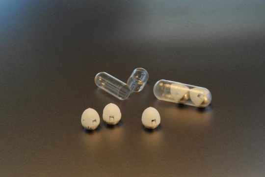
An MIT-led research team has developed a drug capsule that could be used to deliver oral doses of insulin, potentially replacing the injections that people with type 1 diabetes have to give themselves every day.

About the size of a blueberry, the capsule contains a small needle made of compressed insulin, which is injected after the capsule reaches the stomach. In tests in animals, the researchers showed that they could deliver enough insulin to lower blood sugar to levels comparable to those produced by injections given through skin. They also demonstrated that the device can be adapted to deliver other protein drugs.

“We are really hopeful that this new type of capsule could someday help diabetic patients and perhaps anyone who requires therapies that can now only be given by injection or infusion,” says Robert Langer, the David H. Koch Institute Professor, a member of MIT’s Koch Institute for Integrative Cancer Research, and one of the senior authors of the study.

Giovanni Traverso, an assistant professor at Brigham and Women’s Hospital, Harvard Medical School, and a visiting scientist in MIT’s Department of Mechanical Engineering, where he is starting as a faculty member in 2019, is also a senior author of the study. The first author of the paper, which appears in the Feb. 7 issue of Science, is MIT graduate student Alex Abramson. The research team also includes scientists from the pharmaceutical company Novo Nordisk.

Self-orientation

Several years ago, Traverso, Langer, and their colleagues developed a pill coated with many tiny needles that could be used to inject drugs into the lining of the stomach or the small intestine. For the new capsule, the researchers changed the design to have just one needle, allowing them to avoid injecting drugs into the interior of the stomach, where they would be broken down by stomach acids before having any effect.

The tip of the needle is made of nearly 100 percent compressed, freeze-dried insulin, using the same process used to form tablets of medicine. The shaft of the needle, which does not enter the stomach wall, is made from another biodegradable material.

Within the capsule, the needle is attached to a compressed spring that is held in place by a disk made of sugar. When the capsule is swallowed, water in the stomach dissolves the sugar disk, releasing the spring and injecting the needle into the stomach wall.

The stomach wall has no pain receptors, so the researchers believe that patients would not be able to feel the injection. To ensure that the drug is injected into the stomach wall, the researchers designed their system so that no matter how the capsule lands in the stomach, it can orient itself so the needle is in contact with the lining of the stomach.

“As soon as you take it, you want the system to self-right so that you can ensure contact with the tissue,” Traverso says.

The researchers drew their inspiration for the self-orientation feature from a tortoise known as the leopard tortoise. This tortoise, which is found in Africa, has a shell with a high, steep dome, allowing it to right itself if it rolls onto its back. The researchers used computer modeling to come up with a variant of this shape for their capsule, which allows it to reorient itself even in the dynamic environment of the stomach.

“What’s important is that we have the needle in contact with the tissue when it is injected,” Abramson says. “Also, if a person were to move around or the stomach were to growl, the device would not move from its preferred orientation.”

Once the tip of the needle is injected into the stomach wall, the insulin dissolves at a rate that can be controlled by the researchers as the capsule is prepared. In this study, it took about an hour for all of the insulin to be fully released into the bloodstream.

Easier for patients

In tests in pigs, the researchers showed that they could successfully deliver up to 300 micrograms of insulin. More recently, they have been able to increase the dose to 5 milligrams, which is comparable to the amount that a patient with type 1 diabetes would need to inject.

After the capsule releases its contents, it can pass harmlessly through the digestive system. The researchers found no adverse effects from the capsule, which is made from biodegradable polymer and stainless steel components.

Maria José Alonso, a professor of biopharmaceutics and pharmaceutical technology at the University of Santiago de Compostela in Spain, describes the new capsule as a “radically new technology” that could benefit many patients.

“We are not talking about incremental improvements in insulin absorption, which is what most researchers in the field have done so far. This is by far the most realistic and impactful breakthrough technology disclosed until now for oral peptide delivery,” says Alonso, who was not involved in the research.

The MIT team is now continuing to work with Novo Nordisk to further develop the technology and optimize the manufacturing process for the capsules. They believe this type of drug delivery could be useful for any protein drug that normally has to be injected, such as immunosuppressants used to treat rheumatoid arthritis or inflammatory bowel disease. It may also work for nucleic acids such as DNA and RNA.

“Our motivation is to make it easier for patients to take medication, particularly medications that require an injection,” Traverso says. “The classic one is insulin, but there are many others.”

The research was funded by Novo Nordisk, the National Institutes of Health, a National Science Foundation Graduate Research Fellowship, Brigham and Women’s Hospital, a Viking Olaf Bjork Research Scholarship, and the MIT Undergraduate Research Opportunities Program.

Other authors of the paper include Ester Caffarel-Salvador, Minsoo Khang, David Dellal, David Silverstein, Yuan Gao, Morten Revsgaard Frederiksen, Andreas Vegge, Frantisek Hubalek, Jorrit Water, Anders Friderichsen, Johannes Fels, Rikke Kaae Kirk, Cody Cleveland, Joy Collins, Siddartha Tamang, Alison Hayward, Tomas Landh, Stephen Buckley, Niclas Roxhed, and Ulrik Rahbek.

More Sleepless Nights for Toddlers With Autism

by Elizabeth Hlavinka, Staff Writer, MedPage Today

Children with autism spectrum disorder (ASD) were more than twice as likely to have problems sleeping than those without neurodevelopmental disorders, according to a community-based case-control study.

In a sample of nearly 2,000 children ages 2 to 5 years, those with ASD had significantly higher odds of sleep troubles — such as night waking and bedtime resistance — than kids in the general population (adjusted OR 2.37, 95% CI 1.75-3.22), reported Ann Reynolds, MD, of the University of Colorado School of Medicine, and colleagues.

Children with ASD were also more than twice as likely as children with other developmental delays and disorders to have problems sleeping (adjusted OR 2.12, 95% CI 1.57-2.87), they wrote in Pediatrics.

“For all children, lack of sleep can cause increased hyperactivity, inattention, and irritability, but for kids with ASD, poor sleep has been associated with more repetitive behaviors and self injury and so that has an impact on the quality of life for the child,” Reynolds told MedPage Today in an interview in which a media relations representative was present.

Although this study did not examine the cause of this increased risk for sleep disturbances, it’s possible that multiple contributing factors could predispose children with ASD to sleep problems, including obstructive sleep apnea, attention-deficit hyperactivity disorder (ADHD), or behavioral disturbances that interfere with sleep, Reynolds and colleagues noted.

Catherine Lord, PhD, of the David Geffen School of Medicine at UCLA, who wrote an accompanying commentary, said that although the same strategies and medications that improve childhood sleep are typically helpful in children with ASD, certain treatments like sleep training can take longer to change autistic children’s sleep patterns.

“Although medication used for sleep have been shown to be safe and have similar effects in kids with autism, there’s always variation and there will be more variation in kids with autism than in other populations,” Lord told MedPage Today.

Lord stressed the importance of pediatricians and healthcare providers working with parents of children with ASD to develop plans for improving sleep disturbances, particularly because a lack of sleep could further exacerbate their condition.

Reynolds and her team examined data from the Study to Explore Early Development (SEED) study, noting that all children included in this analysis were born from September 2003 to August 2006, completed a developmental evaluation, and had a caregiver interview and a completed Children’s Sleep Habits Questionnaire. Three groups of children were included in the study — those with a research classification of ASD, children with other developmental delays and disorders, and children from the general population.

Notably, a higher score on the 33-item Children’s Sleep Habits Questionnaire indicates more sleep issues, but researchers used two cut-offs for this study. While a total score of 41 differentiates a clinical sample of children with sleep problems, the authors said, they also used a total score cutoff of 48 for a more conservative estimate.

Data was adjusted for mothers’ education, race or ethnicity, and family income, as well as children’s sex, enrollment age, cognitive scores, and prior genetic or neurological diagnoses.

Of the 1,987 children included, those with ASD were more likely to be boys and have lower cognitive scores, and were less likely to have high-income families and non-Hispanic white mothers with a college degree.

Using the standard total score cutoff of 41, the results were “attenuated, but similar” as the more conservative total score cutoff. There were higher odds of sleep problems among children with ASD versus the children in the general population (adjusted OR 1.75, 95% CI 1.27-2.42) and versus children with other developmental delays and disorders (adjusted OR 1.45, 95% CI 1.05-2.00).

There was no difference in sleep patterns observed between children with ASD and children with other developmental delays and disorders in addition to ASD at either cutoff, Reynolds and colleagues noted.

Using the more conservative sleep score cut-off, 47% of parents reported sleep problems in their children with ASD compared to 29% of children with other developmental delays and disorders, and 25% of children in the general population, the authors noted.

Limitations to the data mainly involve limits to the Children’s Sleep Habits Questionnaire. Because it is a parent-completed questionnaire, there is no objective measure of sleep and no clinical assessment of sleep problems, the authors said, meaning “determining the exact prevalence of sleep problems” on the basis of the questionnaire “may be problematic.” In addition, the questionnaire was not designed to make a sleep order diagnosis or address the severity of sleep problems, nor has it been validated for use in young children (ages <4 ) or children with ASD as well, they noted.

Could Diet Sodas Raise Older Women’s Stroke Risk?

By Alan Mozes

HealthDay Reporter

THURSDAY, Feb. 14, 2019 (HealthDay News) — Older women, beware: New research warns that drinking a lot of diet sodas or artificially sweetened fruit juices may increase your risk for stroke.

In a study that tracked nearly 82,000 postmenopausal women, those who drank two or more diet drinks per day saw their overall stroke risk rise by 23 percent, compared with those who consumed diet drinks less than once a week.

Blocked arteries were often the main culprit, with heavy diet drink consumption linked to a 31 percent greater risk for an ischemic stroke, which is triggered by a clot, the study findings showed.

Study author Yasmin Mossavar-Rahmani acknowledged that an “association does not imply causation.” But she stressed that the findings held up even after taking into account the nutritional value of each participant’s overall diet.

So, “we can’t assume these diet drinks are harmless, particularly when consumed at high levels,” Mossavar-Rahmani said.

“The take-home message is that these findings give us pause,” she added. “We need to do more research on why we are seeing these associations. What are the scientific mechanisms? Is there something about the artificial sweeteners, for example, that affect the bacteria in the gut and lead to health issues?”

Mossavar-Rahmani is an associate professor in the department of epidemiology and population health’s division of health promotion and nutrition research at Albert Einstein College of Medicine, in New York City.

The study authors pointed out that the American Heart Association (AHA) has recently underscored the lack of sufficient research into the cardiovascular impact of diet sodas. Until more work is done, the AHA says the jury remains out on whether artificially sweetened beverages do or do not hasten heart disease.

Women in the latest study were between 50 and 79 when they first enrolled in the Women’s Health Initiative trial between 1993 and 1998.

Investigators tracked the general health of all the enrollees for an average of nearly 12 years. During that time — at the three-year mark — all the women were asked to indicate how frequently they consumed diet sodas and diet fruit drinks over a three-month period.

The researchers did not take note of which brands of artificially sweetened drinks the women drank, and so did not know which artificial sweeteners were being consumed.

That said, nearly two-thirds of the women consumed diet sodas or drinks very infrequently, meaning less than once a week or never. Only about 5 percent were found to be “heavy” consumers of artificially sweetened drinks.

After taking into consideration a variety of stroke risk factors — including blood pressure status, smoking history and age — the study team concluded that heavy consumption of diet drinks did appear to be tied to cardiovascular risks in a number of ways.

For example, those women who drank two or more diet beverages a day saw their overall risk for developing heart disease increase by 29 percent. They were also 16 percent more likely to die prematurely from any cause.

Certain groups fared even worse: Among obese women and black women with no history of heart disease or diabetes, a diet drink habit pushed clot-driven stroke risk up by roughly twofold and fourfold, respectively, the researchers reported.

Whether or not the findings would apply to either men or younger women remains unclear, the study authors noted.

The findings were published online Feb. 14 in the journal Stroke.

Lona Sandon is program director of the department of clinical nutrition at the University of Texas Southwestern Medical Center in Dallas.

She agreed that more research is needed to further explore a possible diet drink-heart disease connection. But for now Sandon offered simple advice: diet or regular, sodas offer no nutritional value other than calories.

“If they replace other drinks, such as milk and 100 percent fruit or vegetable drinks, then these women miss out on valuable nutrition for protecting the heart and vascular system,” Sandon warned.

“The nutrition you are missing because you are drinking artificially sweetened beverages instead may be the real problem,” she said.

A group representing the artificial sweetener industry offered another caveat about the findings — that many women who drink diet drinks are already struggling with weight issues.

“It is likely study subjects were already at a greater health risk and chose low-calorie sweetened beverages to manage their calorie and sugar intake as these products are proven safe and beneficial for those managing their weight and blood glucose levels,” said Robert Rankin, president of the Calorie Control Council.

“The contribution of reverse causality, meaning that individuals already at a greater risk of stroke and cardiovascular events chose low-calorie sweetened beverages, is very likely the cause of the associations presented by these researchers,” the council added in a statement.

How music motivates the brain to learn

Humans have been making, listening, and dancing to music since time immemorial, and this art can easily soothe or amplify our emotions. New research explains what “chords” music strikes in the brain, and how it relates to certain cognitive processes, particularly learning.

In recent years, researchers have shown more interest in how listening to music can pragmatically benefit us in numerous ways.

For instance, studies such as this one, which Medical News Today covered last spring, have suggested that people with Alzheimer’s disease who face anxiety and other stressful emotions cope better when they listen to music.

Listening to certain types of music may even alter our perception and change the way we see potential partners, while happy songs can help boost our creativity.

In a new study, the findings of which feature in the journal PNAS, researchers from McGill University in Montreal, Canada have now shown that we can use music to activate the brain’s reward center and motivate learning in an error prediction model.

“Although many authors have proposed that the intense emotions and pleasures of music result from expectancies, predictions, and their outcomes […], direct evidence for this proposition has been lacking,” the researchers write.

The current research finally gets to the bottom of this proposition, using a musical reward learning task and functional MRI to understand how pleasurable music motivates the brain to learn and to strive for its reward.

Music as a reward that ‘supports learning’

The team worked with 20 participants aged between 18 and 27 years, whom they asked to take part in a musical reward experiment. Each person had to choose a combination of colors and directions, and each combination had a different probability of the participant hearing either pleasant music or a dissonant, unpleasant audio track.

After a few attempts, the participants learned which combinations they should choose in order to increase their chances of accessing the enjoyable music reward.

While the volunteers were participating in this task, the researchers used functional MRI to measure their brain activity. Then, using a special algorithm, the investigators calculated the difference between how often the participants expected to receive their reward and the number of times they actually received it.

On further comparing this data with the functional MRI scans, the team found that correct predictions correlated with heightened activity in a brain area called the nucleus accumbens, which previous research has tied to the experience of pleasure when listening to music.

This finding indicates that music is, in itself, a viable reward and one that can provide enough motivation to the brain to learn new information that will allow it to access this source of pleasure more easily.

Furthermore, the participants who found the right combinations and made the correct predictions the most often, which correlated with heightened activity in the nucleus accumbens each time, also made the most learning progress throughout the tasks.

“This study adds to our understanding of how abstract stimuli like music activate the pleasure centers of our brains,” explains study author Benjamin Gold.